Quantum-enabled EPR spectroscopy
Access the inaccessible
Static structures miss the motion that dictates biological function. FATHOM® resolves dynamic behavior.

Structure isn't static.
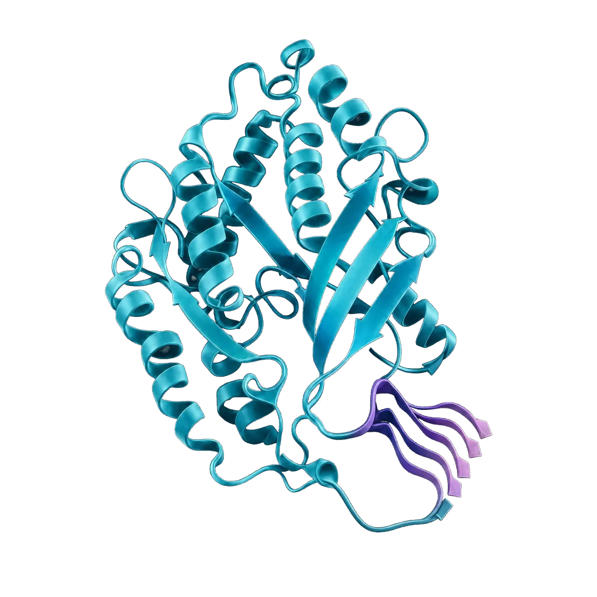
Cryo-EM, NMR, AlphaFold, and X-ray crystallography give you a snapshot. Biological function lives in what the snapshot misses — the loops that flex, the domains that switch, the conformational ensembles that decide how a molecule binds.
Measure the motion.
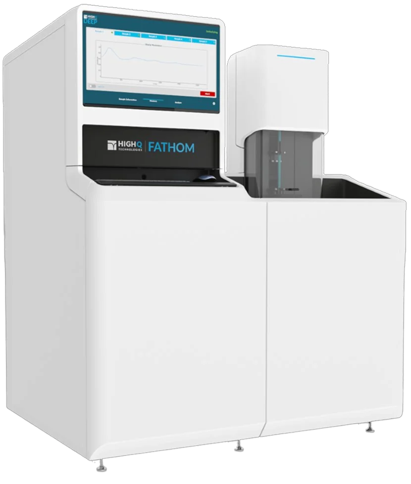
FATHOM resolves nanoscale distance distributions and conformational dynamics on micromolar samples, in under a few hours, on an instrument built for the structural biology lab.
The world's first quantum-enabled EPR spectrometer.
FATHOM measures the protein dynamics that static structural techniques can't resolve — the conformational motion behind binding, signalling, and drug action.
Revolutionizing EPR with Quantum Sensing Technology
Next-generation quantum sensing enhances EPR with greater sensitivity, faster acquisition, and simplified operation. Automated workflows and an intuitive interface enable high-throughput analysis, reducing experiment times from weeks to days.
Superconducting Quantum Sensing
New superconducting quantum-sensing resonator design enables accessible scientific discoveries and higher throughput than classical EPR.
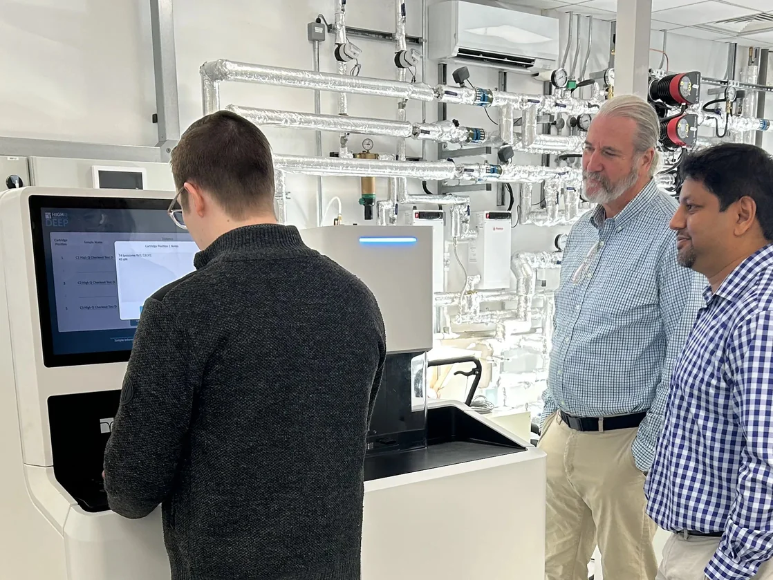
Easy Automated Operation
Load-and-go automated reporting, multi-sample handling and an intuitive touchscreen interface designed for technician-level users.
Short Acquisition Times
Increase in sensitivity, speed and stability reduces EPR acquisition time from weeks to days.

Born from quantum research. Built to transform drug discovery.
High-Q Technologies emerged from the Institute for Quantum Computing at the University of Waterloo. Backed by Quantum Valley Investments and guided by scientific advisors from MIT, we are building the measurement infrastructure for the next generation of structural biology.
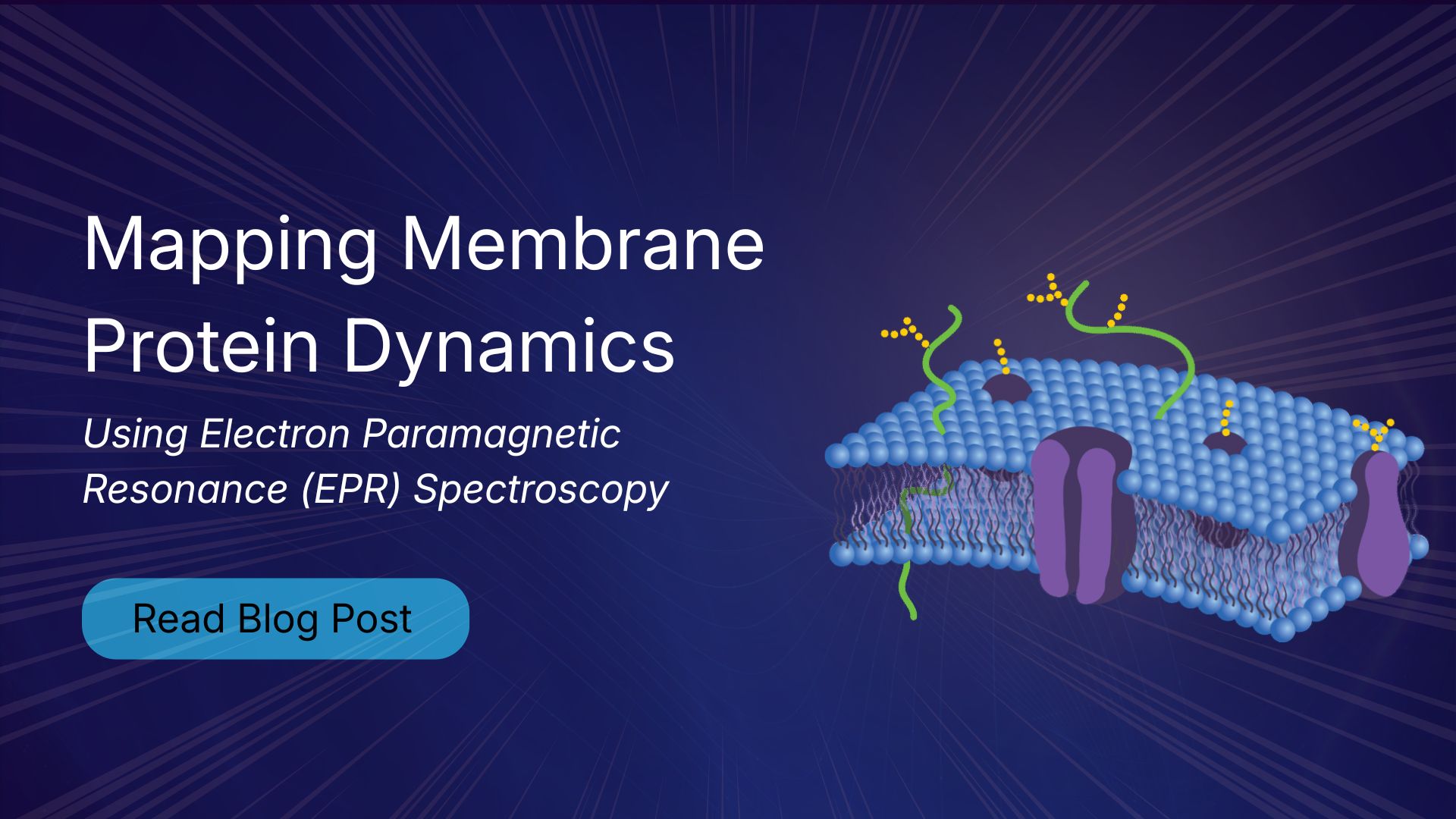
Electron paramagnetic resonance (EPR) spectroscopy is one of the most effective methods for studying membrane protein structure and dynamics. Unlike X-ray crystallography and cryo-electron microscopy, which provide static structural snapshots, EPR measures conformational ensembles and molecular motions under native-like conditions. This makes it particularly valuable for investigating GPCRs, ion channels, and membrane transporters involved in drug discovery.

The Rocky Mountain Conference on Magnetic Resonance remains one of the premier gatherings for the EPR community. This year, High Q Technologies will share new advances in automated EPR spectroscopy with the FATHOM system, highlighting improved sensitivity, phase stability, and applications in structural biology and challenging membrane protein research.

As structural biology/drug discovery moves toward ensemble-based views of proteins, Electron Paramagnetic Resonance (EPR) spectroscopy is becoming one of the most important techniques for studying biomolecular dynamics. EPR studies are increasingly being used to complement existing structural data from techniques like Cryo-EM, NMR, or X-ray Crystallography, providing dynamic information that can be difficult to obtain otherwise. In this post, we will take a look at Electron Paramagnetic Resonance spectroscopy (EPR), and why it keeps showing up in protein dynamics studies.