Protein dimerization is one of the most widespread and functionally important forms of protein–protein interaction. Proteins rarely act in isolation in a biological environment; instead, they associate as homodimers, heterodimers or higher-order oligomers to form functional assemblies. This self-assembly occurs in every cellular compartment, from membranes to the nucleus and cytosol, and regulates processes ranging from enzymatic activation and signal transduction to pathogenic pathways. A dimer observed in a crystal structure, however, may represent either a genuine biological assembly or a contact created by crystal packing. A crystal structure provides detailed information about molecular architecture, but it does not by itself establish whether two molecules associate under relevant solution conditions, how strong that interaction is, or whether the subunits retain the same arrangement outside the lattice. Distinguishing between these possibilities is therefore an important task in structural biology, biophysics and pharmaceutical research, where protein–protein interactions can influence function, stability and the binding of potential drug candidates.
How to Validate a Protein Dimer
Determining whether a crystallographic dimer is real can be approached through three related questions. The first is whether the dimer forms in solution, that is, whether the protein exists as a monomer, a dimer or a mixture of oligomeric states under the conditions of interest. The second is whether the dimer adopts the proposed structure, meaning whether experimental measurements support the relative arrangement of the subunits in the structural model. The third is how stable or dynamic the interaction is, and whether the dimer remains associated, dissociates or changes conformation in response to concentration, ligands or environmental conditions. Analytical ultracentrifugation (AUC), size-exclusion chromatography coupled to multi-angle light scattering (SEC-MALS), native mass spectrometry (native MS) and double electron–electron resonance (DEER) spectroscopy provide complementary ways of addressing these questions. Because each technique measures a different physical property, no single method answers all three. A technique that establishes the presence of a dimer does not necessarily reveal its geometry, while a structural measurement does not automatically quantify the proportion of protein present as a dimer.
What Is Protein Dimerization?
Protein dimerization occurs when two protein molecules associate to form a complex known as a dimer. When both subunits are identical, the complex is a homodimer; when they differ, it is a heterodimer. Some proteins form stable dimers, whereas others undergo reversible association, with their oligomeric state depending on protein concentration, temperature, buffer composition, ligand binding and other environmental factors. For a reversible homodimer, the equilibrium between monomer (M) and dimer (D) can be represented as:
2M ⇌ D
The dissociation constant is:
Kᴅ = [M]² / [D]
Here, the brackets indicate molar concentrations at equilibrium, so K_D has units of concentration. A lower K_D generally indicates stronger association, while a higher K_D indicates weaker association under the conditions measured. Importantly, the proportion of protein present as a dimer also depends on the total protein concentration. A protein may therefore be predominantly dimeric at the high concentrations used for crystallization yet contain a substantial monomer fraction after dilution. Observing a dimer at one concentration does not, on its own, establish that it remains dimeric under other conditions.
Methods for determine whether a protein dimer is real in solution
Several analytical methods can establish whether a protein exists as a monomer or dimer in solution. They differ in the physical properties they measure, the information they provide and their sensitivity to sample conditions.
Analytical ultracentrifugation (AUC) measures how proteins behave under centrifugal force. Sedimentation-velocity experiments characterize the distribution of species in a sample and can identify heterogeneity, while sedimentation-equilibrium experiments are particularly useful for investigating reversible self-association and estimating monomer–dimer dissociation constants. When dimerization is concentration-dependent, measurements across a range of concentrations can help distinguish between alternative association models. AUC is therefore well suited to characterizing self-association and quantifying the strength of a reversible monomer–dimer equilibrium.
Size-exclusion chromatography coupled to multi-angle light scattering (SEC-MALS) estimates the molar mass of protein species as they elute from a column, allowing species consistent with monomers, dimers or larger assemblies to be distinguished. It is a practical choice for characterizing purified proteins and assessing sample heterogeneity or higher-order assembly. However, the chromatographic step separates and dilutes the sample, so weak, reversible interactions may shift during the run, and the observed distribution may not reflect the equilibrium at the original concentration. For weak interactions, an independent method is advisable.
Native mass spectrometry (native MS) provides information about the mass and stoichiometry of noncovalent complexes and can resolve multiple oligomeric species, making it valuable for determining complex composition. Sample preparation, ionization and complex stability during measurement can influence which species are detected, so native MS results are best interpreted as complementary evidence within the context of the experimental conditions.
Electron Paramagnetic Resonance as a Tool to Validate Dimer Structure
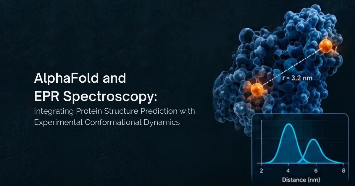
Double electron–electron resonance (DEER), also known as pulsed dipolar EPR spectroscopy or PELDOR, measures dipolar interactions between paramagnetic spin labels. When combined with site-directed spin labelling, it provides distance distributions between selected positions on a protein. These distances can be compared with predictions from a crystal structure or alternative models, making DEER particularly valuable when the research question concerns the relative arrangement of subunits rather than simply their molecular mass.
DEER also has important limitations. Spin-label flexibility, experimental uncertainty and the presence of multiple conformations can affect interpretation, and a single distance rarely establishes a unique structure. It is equally important to distinguish structural distance measurements from measurements of oligomeric populations. Conventional distance-distribution analysis does not automatically determine the fraction of protein present as a dimer or its dissociation constant, although specialized approaches such as inversion-modulated DEER can resolve monomer–dimer populations under suitable conditions.
The choice between DEER and methods such as AUC or SEC-MALS therefore depends on the question being asked. Whether a protein forms a dimer in solution can be addressed by AUC, SEC-MALS or native MS, and the strength of a reversible equilibrium is best estimated from concentration-dependent AUC. Whether a crystallographic dimer has the correct subunit geometry, or how that geometry responds to ligands or subunit exchange, is where DEER contributes most. For many systems, the most informative strategy combines a method that characterizes oligomeric state with one that tests the proposed structure.
A DEER experiment designed to test a proposed dimer begins with a structural hypothesis. Labelling positions are first selected so that their distances distinguish the proposed dimer from plausible alternative arrangements, with multiple independent distances generally more informative than one. Spin labels are then introduced, most commonly through engineered cysteine residues and suitable label chemistry. The resulting distance distributions are measured, and their width and shape can reflect structural heterogeneity or distinct conformational states. Finally, these distributions are compared with distances predicted from structural models, taking spin-label flexibility and uncertainty into account.
Agreement between experimental and predicted distributions supports the compatibility of a model with the data. Even several matching distances, however, do not independently establish that a dimer is biologically relevant; that conclusion also requires evidence that the assembly forms under the conditions of interest.
DEER EPR Studies of Protein Dimerization
DEER has been applied to questions that extend well beyond measuring a single structural distance. Four published studies illustrate distinct applications: environment-dependent association, monomer–dimer population analysis, ligand-induced conformational change and subunit-exchange kinetics.
Dimer Stability Inside Cells
Most dimer characterization is carried out on purified protein in buffer, yet the cellular environment is crowded and chemically complex. To investigate how this environment affects association of the homodimeric BIR1 domain of XIAP, Yang et al. (2020) used DEER with Gd(III) spin labels, which are relatively stable in the reducing intracellular environment. They found that the dimer was less stable in cells than in solution or dilute cell lysate, with a correspondingly higher dissociation constant, and further examined the effects of salt and molecular crowding agents. These results show that a dimer characterized in buffer may not have the same stability in its native environment.
Resolving Monomer–Dimer Populations
Conventional DEER analysis reports distances rather than populations. Schmidt, Ghirlando, Baber and Clore (2016) demonstrated that inversion-modulated DEER, which analyses modulation depth as a function of the pump-pulse flip angle, can resolve monomer and dimer populations directly. Applied to the p66 subunit of HIV-1 reverse transcriptase, the approach revealed a monomer–dimer equilibrium whose relative populations were affected by glycerol concentration. Quantifying populations therefore requires an experimental design that explicitly supports it, and the resulting values must be interpreted under the conditions of the EPR sample.
Ligand-Induced Changes in Dimer Geometry
tRNA-guanine transglycosylase (TGT) is a homodimeric enzyme, which makes it a useful system for asking whether ligand binding alters the arrangement of subunits within a dimer. Nguyen et al. (2021) used site-directed spin labelling and PELDOR/DEER spectroscopy to measure distances across the dimer interface and revealed a ligand-dependent equilibrium between different dimer conformations, including a twisted state. The authors proposed a dissociation–association mechanism for formation of the twisted dimer upon ligand binding. These results show that a protein can remain dimeric while its subunits change their relative arrangement, and that DEER can provide the distance constraints needed to evaluate models of each state.
Measuring Subunit-Exchange Kinetics
A crystal structure provides a static snapshot and cannot reveal how quickly subunits exchange. Yang et al. (2013) combined DEER with NMR paramagnetic relaxation enhancement (PRE) to study subunit exchange in the Dsy0195 homodimer from Desulfitobacterium hafniense. Their analysis yielded a subunit-dissociation rate constant of 0.037 ± 0.005 min⁻¹, corresponding to a dissociation half-life of approximately 18.7 minutes, in agreement with independent PRE measurements. With suitable experimental design and kinetic analysis, DEER can therefore contribute to measurements of protein-complex dynamics.
Combining Methods to Validate a Protein Dimer
Determining whether a crystallographic dimer is real requires more than identifying a plausible interface. It requires evidence that the protein associates in solution, that the proposed subunit arrangement is consistent with experimental data and, where relevant, an understanding of the interaction’s stability and dynamics. AUC, SEC-MALS and native MS characterize protein assembly, while DEER provides distance constraints for evaluating structural models and, with appropriate experimental design, can contribute to studies of oligomeric populations, conformational change and subunit-exchange kinetics.
The most informative strategy is to match each method to the question being asked and to combine independent lines of evidence. This approach distinguishes a crystal-packing contact from a supported solution-state assembly without overstating what any single experiment can establish, and provides a sound structural foundation for downstream work in structural biology and drug discovery.
Frequently Asked Questions
How should spin labels be placed to test a symmetric homodimer interface?
In a symmetric homodimer, a single labelled site per subunit produces one intersubunit distance across the symmetry axis, so each single-cysteine mutant yields one distance constraint. Sites should be chosen so that the competing models predict clearly different distances at that position. Positions very close to the symmetry axis give distances too short for reliable DEER measurement, while positions far apart may exceed the accessible range, which depends on sample relaxation and deuteration. Sites should also avoid the interface itself where labelling could disrupt association.
How many labelling sites are usually needed?
The answer depends on whether the goal is to test an existing model or to determine the dimer arrangement. Testing whether data are compatible with one crystal contact rather than another can be achieved with a small number of well-chosen sites, provided the alternatives predict distinct distances. Determining an arrangement requires more.
How can a crystallization artefact be distinguished from a weak, concentration-dependent dimer?
Both can appear as a monomer in dilute solution, so a single measurement at one concentration cannot separate them. A genuine weak dimer shows reversible, concentration-dependent association that can be fitted to a monomer–dimer model with a measurable dissociation constant, for example by sedimentation-equilibrium AUC across a concentration series. A crystal-packing contact, by contrast, shows no significant association even at high concentrations. DEER modulation depth also reports on the fraction of dimer present, so measurements across concentrations can contribute the same information, as in the BIR1 study.
References and further reading
- Yang et al. (2020). In-cell destabilization of a homodimeric protein complex detected by DEER spectroscopy. PNAS, 117(34), 20566–20575.
- Schmidt, T., Ghirlando, R., Baber, J. and Clore, G. M. (2016). Quantitative Resolution of Monomer-Dimer Populations by Inversion Modulated DEER EPR Spectroscopy. ChemPhysChem, 17(19), 2987–2991.
- Nguyen et al. (2021). Unraveling a Ligand-Induced Twist of a Homodimeric Enzyme by Pulsed Electron–Electron Double Resonance. Angew. Chem. Int. Ed., 60(43), 23419–23426.
- Yang et al. (2013). Measurement of Rate Constants for Homodimer Subunit Exchange Using Double Electron–Electron Resonance and Paramagnetic Relaxation Enhancements. J. Biomol. NMR, 55(1), 47–58.
- Jeschke, G. (2012). DEER Distance Measurements on Proteins. Annu. Rev. Phys. Chem., 63, 419–446.
- Tessmer and Stoll (2025). Protein Modeling with DEER Spectroscopy. Annu. Rev. Biophys., 54, 35–57.
- Kerry, P. S., Turkington, H. L., Ackermann, K., Jameison, S. A. and Bode, B. E. (2014). Analysis of Influenza A Virus NS1 Dimer Interfaces in Solution by Pulse EPR Distance Measurements. J. Phys. Chem. B, 118(37), 10882–10888.
- Hale, B. G., Barclay, W. S., Randall, R. E. and Russell, R. J. (2008). Structure of an avian influenza A virus NS1 protein effector domain. Virology, 378, 1–5.
- Hilger et al. (2007). High-Resolution Structure of a Na⁺/H⁺ Antiporter Dimer Obtained by Pulsed Electron Paramagnetic Resonance Distance Measurements. Biophysical Journal.