Insights from Austin
The latest insights on EPR technology trends, customer successes and industry best practises

Protein dimerization is one of the most widespread and functionally important forms of protein–protein interaction. Proteins rarely act in isolation in a biological environment; instead, they associate as homodimers, heterodimers or higher-order oligomers to form functional assemblies. This self-assembly occurs in every cellular compartment, from membranes to the nucleus and cytosol, and regulates processes ranging from enzymatic activation and signal transduction to pathogenic pathways. A dimer observed in a crystal structure, however, may represent either a genuine biological assembly or a contact created by crystal packing. A crystal structure provides detailed information about molecular architecture, but it does not by itself establish whether two molecules associate under relevant solution conditions, how strong that interaction is, or whether the subunits retain the same arrangement outside the lattice. Distinguishing between these possibilities is therefore an important task in structural biology, biophysics and pharmaceutical research, where protein–protein interactions can influence function, stability and the binding of potential drug candidates.
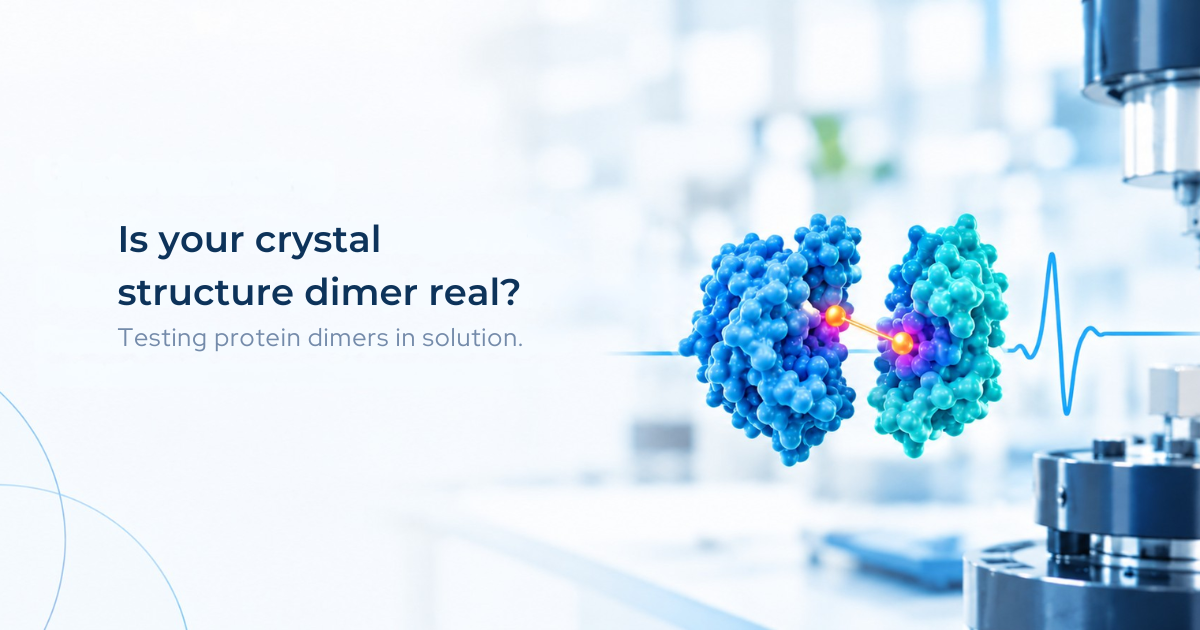
Protein dimerization occurs when two protein molecules associate to form a dimer. A crystal structure can reveal how two protein molecules fit together, identifying an interface that may influence protein function or drug discovery. However, a dimer observed in a crystal does not necessarily represent the dominant assembly in solution. Crystal packing can stabilize contacts that are weak, transient, or absent under physiologically relevant conditions. For pharmaceutical researchers, this distinction matters because a proposed dimer interface may form a binding site, regulate protein activity, or provide a target for small molecules. Before using the structure to guide drug design, researchers need to establish whether the protein associates in solution and whether the arrangement of its subunits is consistent with the proposed model.
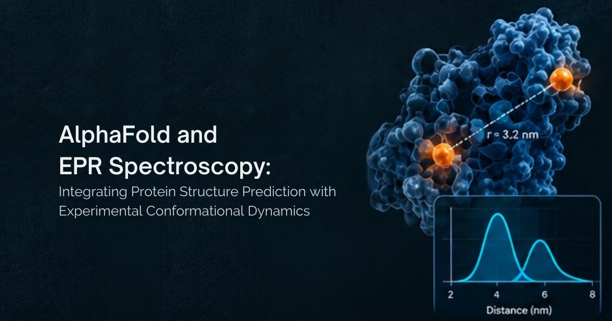
Few tools have changed structural biology as quickly as AlphaFold2. Within a few years of its release, AlphaFold2 had become an important starting point for structural studies of many proteins. However, transporters, receptors, channels and many enzymes function through conformational changes, and ligand binding can shift proteins between different structural states. A single predicted model, however confident, represents one structural state rather than the full range of conformations a protein may populate. This is where Electron Paramagnetic Resonance (EPR) spectroscopy comes in.
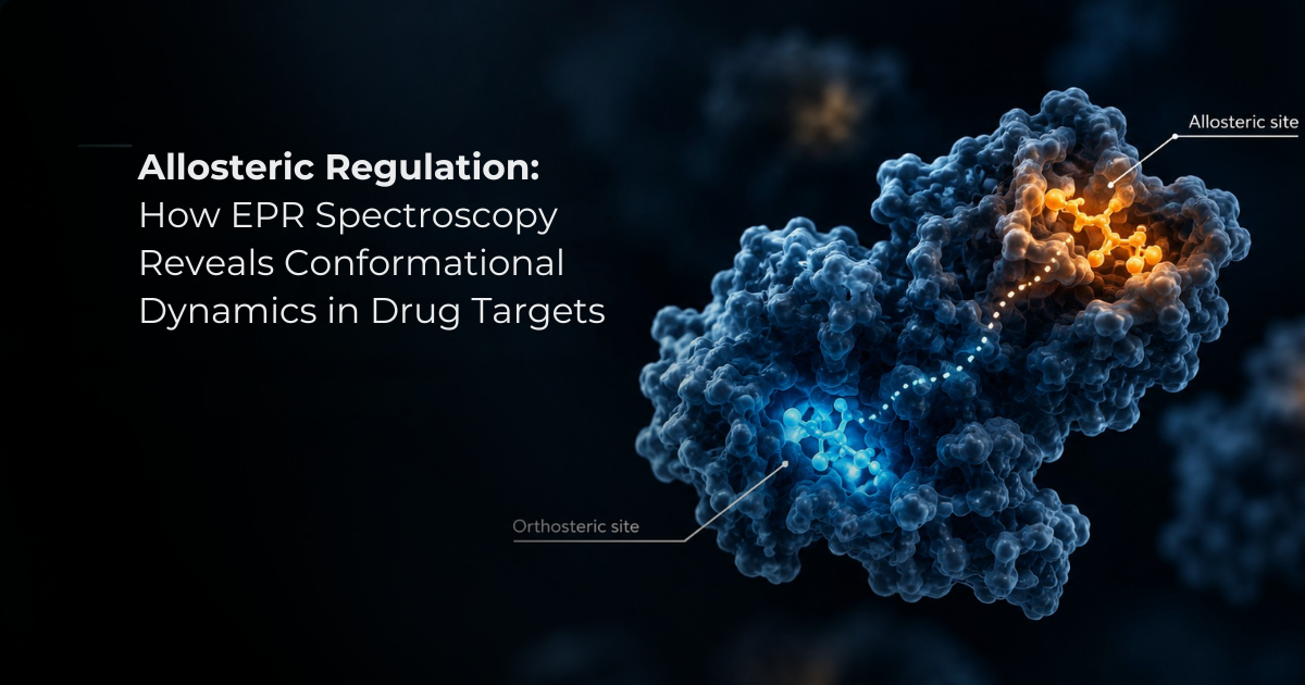
Allosteric regulation is a fundamental mechanism of cellular control, enabling proteins to respond to signals at one site and translate them into changes in activity at another, often distant, site. Proteins regulated in this way, known as allosteric proteins, are involved in a wide range of biological processes, including enzyme regulation, signal transduction, cell adhesion and transcription.
Electron paramagnetic resonance spectroscopy is a powerful way to study materials, molecules, and systems with unpaired electrons. It is often called EPR, ESR, electron spin resonance spectroscopy, or spin resonance spectroscopy.
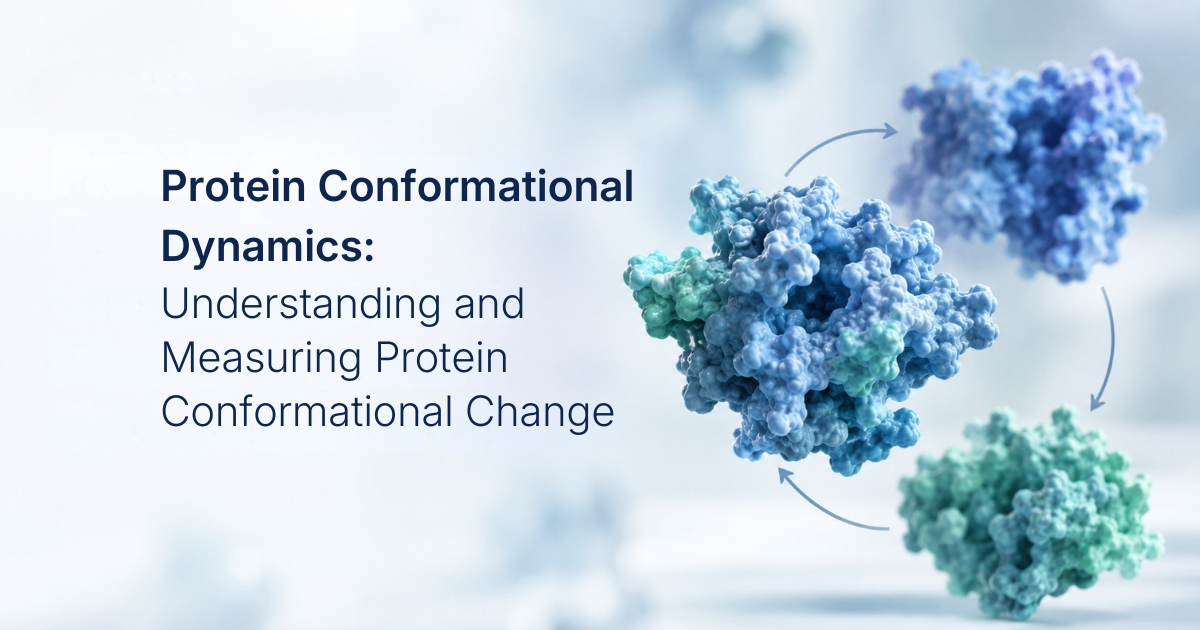
A protein conformational change is often described as a transition from one structural state to another: open to closed, inactive to active, or apo to ligand-bound. That language is useful for describing structural differences, but it can obscure the physical problem that determines how a protein behaves in solution.
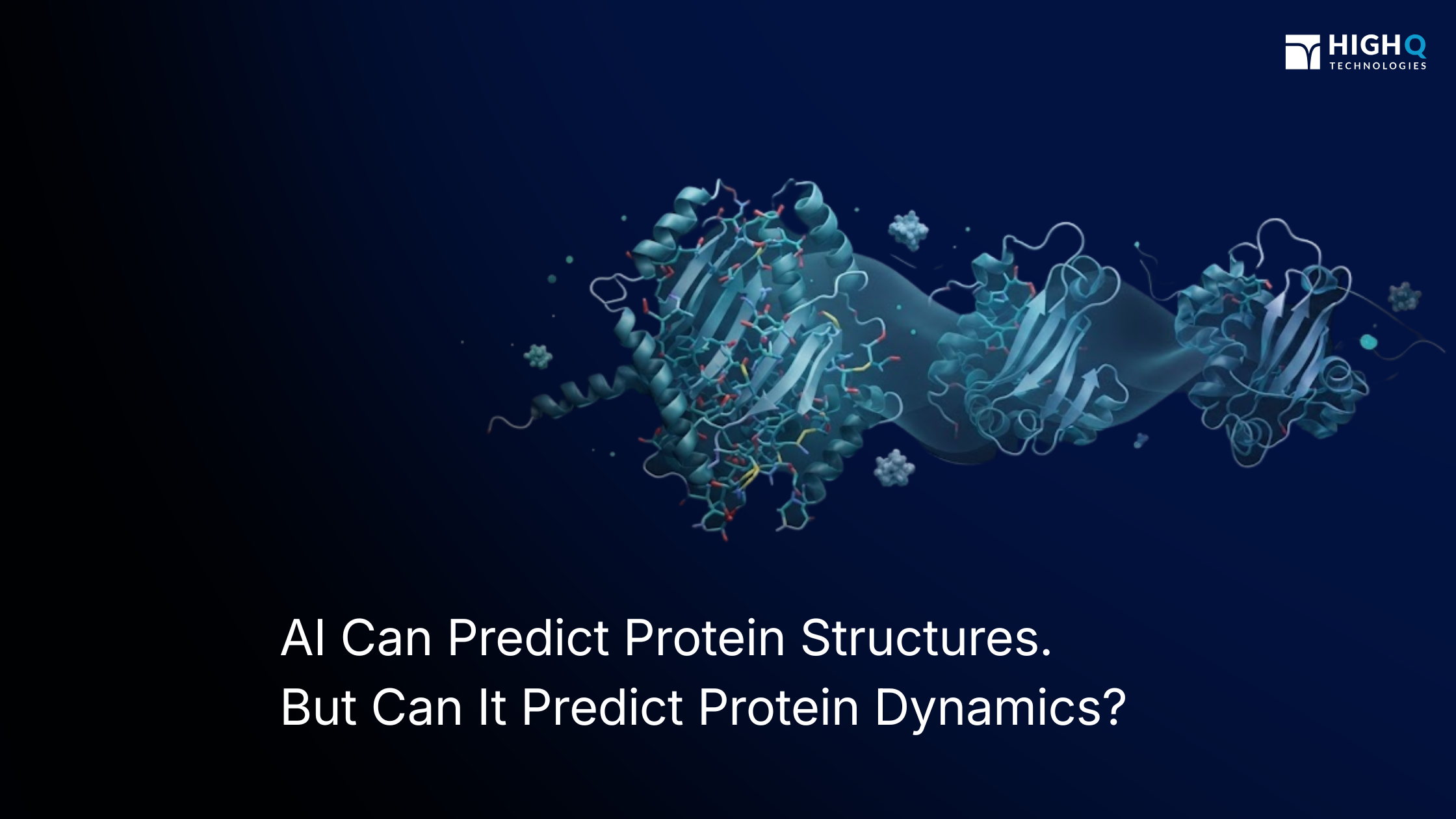
Artificial intelligence has changed the starting point for structural biology. Protein structure prediction tools such as AlphaFold can generate highly accurate structural models for many proteins, including proteins for which experimentally determined structures are unavailable.

Structure-based drug design uses the three-dimensional structure of a target protein to identify functional binding sites and design drug molecules that complement their shape, size, and electrostatic properties. This approach has enabled major advances in medicine, including HIV protease inhibitors, antibiotics, and targeted cancer therapies such as imatinib.
.png)
A high-resolution cryo-EM map can still leave important regions unresolved. Here's what missing density may and may not tell you about protein structure, flexibility, and conformational heterogeneity.
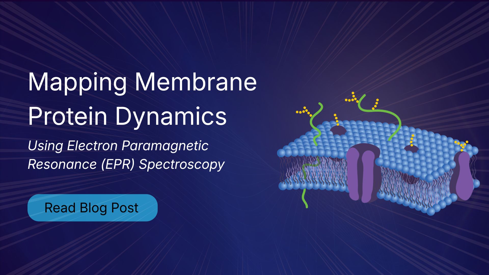
Electron paramagnetic resonance (EPR) spectroscopy is one of the most effective methods for studying membrane protein structure and dynamics. Unlike X-ray crystallography and cryo-electron microscopy, which provide static structural snapshots, EPR measures conformational ensembles and molecular motions under native-like conditions. This makes it particularly valuable for investigating GPCRs, ion channels, and membrane transporters involved in drug discovery.

The Rocky Mountain Conference on Magnetic Resonance remains one of the premier gatherings for the EPR community. This year, High Q Technologies will share new advances in automated EPR spectroscopy with the FATHOM system, highlighting improved sensitivity, phase stability, and applications in structural biology and challenging membrane protein research.

As structural biology/drug discovery moves toward ensemble-based views of proteins, Electron Paramagnetic Resonance (EPR) spectroscopy is becoming one of the most important techniques for studying biomolecular dynamics. EPR studies are increasingly being used to complement existing structural data from techniques like Cryo-EM, NMR, or X-ray Crystallography, providing dynamic information that can be difficult to obtain otherwise. In this post, we will take a look at Electron Paramagnetic Resonance spectroscopy (EPR), and why it keeps showing up in protein dynamics studies.

Techniques like Nuclear Magnetic Resonance (NMR) spectroscopy have long been a cornerstone of structural biology, providing atomic-level insight into protein structure and dynamics in solution. NMR remains one of the most powerful tools for studying folded protein domains, as well as local, site-specific dynamics in partially disordered and flexible regions.

As the Biophysical Society Annual Meeting approaches, it is a good time to reflect on the progress the community has made in the field in years past as well as aspire to continue pushing boundaries as we look toward the future.

Drug discovery cannot happen in a vacuum – it requires a constant influx of new ideas, information, and technology to advance. During my recent attendance at Discovery on Target, there was a consistent underlying message that despite the proprietary nature of an organization’s approach, drug discovery is a group effort. With collaboration a key theme throughout the conference, I want to expand on some of the more salient talking points, and how I believe High Q and the EPR community can contribute to the conversation.

Advancements in science are often spurred by the ‘new.’ Be it new technology, new techniques, or new ideas, novel abilities will beget novel outcomes. The field of EPR has seen this effect throughout its history – beginning with the discovery of the phenomenon itself, and driven forward by breakthroughs such as the development of site-directed spin labeling and distance measurement techniques that enable structural studies on biomolecules; membrane mimetics and in-cell/in-vivo methods to capture physiologically relevant data; in-silico modeling, simulation, and analyses packages; and improvements in hardware, resonators, and pulse-shaping. It is a never-ending upward spiral of new discoveries prompting more questions, requiring new tools to investigate. During my recent visit to the Rocky Mountain Conference on Magnetic Resonance in Denver, several talks emphasized just how far this field has come, how it has benefitted from such advancements, and what new tools could push it even further.