Allosteric regulation is a fundamental mechanism of cellular control, enabling proteins to respond to signals at one site and translate them into changes in activity at another, often distant, site. Proteins regulated in this way, known as allosteric proteins, are involved in a wide range of biological processes, including enzyme regulation, signal transduction, cell adhesion and transcription. Through these long-range interactions, allostery allows protein activity to be adjusted in response to changes in the cellular environment and contributes to the maintenance of cellular homeostasis. Dysregulation of allosteric mechanisms has been implicated in numerous human diseases.
The concept of allostery emerged from early studies of protein cooperativity and was formalized through the pioneering work of Monod, Wyman, and Changeux and of Koshland, Némethy, and Filmer in the 1960s. While these classical models linked allostery to ligand-induced conformational changes, contemporary views increasingly describe proteins as dynamic ensembles of interconverting conformational states. In this framework, ligand binding, mutations, or post-translational modifications alter the populations and energetics of these states, thereby reshaping the protein energy landscape and modulating function through thermodynamic coupling between distant regions.
The shift from static structural models toward ensemble-based descriptions of protein function has also influenced modern drug discovery strategies.Traditional drug discovery has primarily focused on orthosteric sites that bind endogenous ligands or substrates. These sites are often conserved across related proteins, which can make achieving the desired selectivity difficult. In contrast, allosteric sites often exhibit greater structural and functional diversity, providing additional opportunities for selective modulation of protein activity. Allosteric modulators can regulate function through mechanisms distinct from direct competition at the orthosteric site, including altering conformational equilibria, signaling properties, or protein–protein interactions. Consequently, allosteric modulation has emerged as an important strategy in therapeutic development and has expanded the range of proteins amenable to pharmacological intervention.
Despite significant advances in structural biology, elucidating the molecular basis of allosteric regulation remains challenging. Proteins are inherently dynamic molecules that populate multiple conformational states across a broad range of spatial and temporal scales. Although X-ray crystallography, cryo-electron microscopy, nuclear magnetic resonance spectroscopy, and computational approaches have provided important insights into allosteric mechanisms, no single technique can fully capture the conformational heterogeneity and dynamic processes that underlie allosteric communication.
Methods capable of directly probing protein dynamics and conformational heterogeneity are therefore essential for understanding allosteric mechanisms and linking structural changes to functional outcomes. Among these, electron paramagnetic resonance (EPR) spectroscopy , particularly double electron–electron resonance (DEER), provides a powerful means of characterizing conformational changes and distance distributions within proteins.This review discusses the molecular basis of allosteric regulation, its implications for drug discovery, and the contribution of EPR spectroscopy to the characterization of allosteric protein dynamics.
Impact of Allosteric Modulation in Drug Discovery
The impact of allosteric modulation on drug discovery extends beyond conventional small-molecule approaches. Allosteric strategies have been applied across diverse target classes, including GPCRs, ion channels, kinases, protein–protein interactions, and nuclear hormone receptors, with multiple allosteric modulators progressing into clinical development or receiving regulatory approval.
Allosteric ligands can produce different pharmacological effects depending on their mechanism of action. These include positive allosteric modulators (PAMs), which potentiate agonist-mediated receptor responses, and negative allosteric modulators (NAMs), which noncompetitively reduce activity. Silent or neutral allosteric modulators (SAMs or NALs) bind to allosteric sites without affecting the response to the orthosteric ligand.
These mechanisms have translated into approved therapies across different target classes. For example, maraviroc is a negative allosteric modulator of the CCR5 receptor, while cobimetinib is an allosteric inhibitor of MEK1/2. Other examples include the allosteric IDH2 inhibitor enasidenib, the IDH1 inhibitor ivosidenib, and the GABAA_A receptor PAM brexanolone.
The field has also expanded beyond traditional small molecules to include emerging modalities such as covalent allosteric inhibitors, protein degraders, peptides and peptidomimetics, and monoclonal antibodies. These developments have broadened the application of allosteric drug discovery across different therapeutic targets and chemical modalities. Despite these advances, identifying suitable allosteric binding sites remains an important challenge, particularly during early lead-generation efforts. Thanks to advances in structural biology, X-ray crystallography, cryo-EM, EPR spectroscopy and screening approaches have provided new opportunities to identify and characterize allosteric sites.
Experimental Approaches to Studying Allostery
X-ray Crystallography
X-ray crystallography is one of the most commonly used methods for studying allostery. It provides detailed structural information about proteins and allows researchers to compare different functional states of the same protein. For example, comparing a protein structure before and after binding of an allosteric ligand can reveal changes in the active site or in other regions of the protein. These structural differences can help explain how a change at one site is transmitted to a distant site within the protein. However, X-ray crystallography mainly provides static structural snapshots and therefore does not directly capture the dynamic movements of proteins. In addition, interactions between the protein and the surrounding crystal lattice may affect its conformation and could result in structural changes that do not fully represent the protein in solution.
NMR spectroscopy
Nuclear magnetic resonance (NMR) spectroscopy is widely used to study both the structure and dynamics of proteins involved in allosteric regulation. One of its main advantages is its ability to detect relatively small structural changes and changes in the populations of different conformational states. NMR can also provide information about protein motions over a wide range of timescales, from picoseconds to seconds. This makes it particularly useful for studying allostery as a dynamic process. For example, NMR can reveal changes in conformational equilibria that occur when a ligand binds to an allosteric site. However, conventional NMR is generally easier to apply to smaller proteins, with proteins below approximately 40 kDa being more suitable for many applications. Larger proteins can produce more complex spectra and broader signals, making structural and dynamic analysis more challenging.
Mass Spectrometry
Native mass spectrometry (native MS) is another useful approach for investigating allosteric interactions. In native MS, proteins and protein complexes are analyzed under conditions that help preserve their noncovalent interactions. This allows researchers to determine the composition of protein complexes and examine how many ligand molecules are bound to a protein. The simultaneous detection of different ligand-bound species can provide information about binding equilibria and how ligand binding affects different molecular states. However, native MS provides less direct structural information than techniques such as X-ray crystallography, NMR, or EPR. Closely related conformational states may also be difficult to distinguish if they have similar mass-to-charge properties. In addition, some interactions or conformations present in solution may be altered during the transition to the gas phase, which needs to be considered when interpreting the results.
Computational methods
Computational methods have become important in the study of allostery as more experimental protein structures and other types of structural data have become available. These approaches can complement experimental techniques used in structure-based drug discovery by providing information about protein flexibility, conformational changes, communication between distant regions, and potential allosteric binding sites. Combining experimental data with computational analysis can also help make the search for allosteric modulators more systematic and less dependent on chance.
Despite these advances, no single computational method can fully describe all types of allosteric behavior. One of the main challenges in the computational study of allostery is identifying suitable allosteric binding sites. These sites are often difficult to detect because they may be hidden or only temporarily available. Such cryptic binding pockets may become accessible when a protein moves into a less-populated conformational state.
EPR spectroscopy
Electron paramagnetic resonance (EPR) spectroscopy is another method for studying protein structure and dynamics. Using site-directed spin labeling, in which a paramagnetic probe is attached to a specific position on the protein, pulsed EPR methods such as double electron–electron resonance (DEER) can measure distances between pairs of spin labels. These measurements provide distance distributions that can be used to identify different conformational states and monitor changes caused by ligand binding or other perturbations. EPR is particularly useful for studying conformational flexibility and heterogeneity, especially when several states exist at the same time.
Learn more about Electron Paramagnetic Resonance Spectroscopy:What is EPR Spectroscopy? Principles, Spin Labeling, and DEER in Proteins - High Q Technologies Inc. | HighQ Technologies
EPR Spectroscopy in the Study of Allosteric Regulation
Electron paramagnetic resonance (EPR) spectroscopy, particularly double electron–electron resonance (DEER), provides a powerful approach for studying the conformational changes underlying allosteric regulation. Most proteins do not contain suitable endogenous paramagnetic centers, hence, protein conformational studies employ site-directed spin labeling (SDSL), in which paramagnetic probes such as nitroxide labels are introduced at selected residues. DEER measures the dipolar interaction between two spin labels and provides a distribution of interspin distances, allowing conformational heterogeneity and changes in the relative populations of different states to be characterized.
Through the comparison of distance distributions in the absence and presence of orthosteric or allosteric ligands, DEER can reveal ligand-induced shifts in conformational equilibria and thereby provide insight into allosteric regulation. In drug discovery, this approach can support mechanism-of-action studies by determining how ligands alter protein conformational ensembles, and can aid hit and lead characterization by comparing whether chemically distinct compounds stabilize similar or different conformational states. Measurements at multiple labeling sites can further identify regions that respond to ligand binding and provide distance constraints that can be integrated with structural models and complementary biochemical or biophysical data to test proposed mechanisms of allosteric regulation.
Case studies:
Case study 1: Cyclin-dependent kinases (CDKs)
Cyclin-dependent kinases (CDKs) are key regulators of the eukaryotic cell cycle.To become fully active, CDKs require both regulatory phosphorylation and binding to a cyclin, a regulatory protein that binds to and helps activate the kinase. Cdk2 is a CDK involved in controlling the transition from the G1 phase to the S phase of the cell cycle. Although Cdk2 activation requires both regulatory phosphorylation and binding of a cognate cyclin, the study aimed to determine how these regulatory signals are allosterically coupled through changes in the kinase’s conformational states.
DEER spectroscopy was used to monitor conformational changes in Cdk2 in solution.Spin labels were introduced at selected positions on the protein, allowing distances between different regions to be measured. One set of measurements monitored the activation loop (A-loop), a flexible region that helps regulate access to the kinase active site while another monitored the αC-helix, a structural element involved in kinase activation. Changes in these distances allowed the researchers to determine how the populations of different conformational states changed in response to cyclin binding, phosphorylation, and inhibitor binding.
DEER measurements showed that Cdk2 exists as a mixture of different conformational states.Cyclin A binding shifted the population toward the A-loop-out state in which the activation loop moves away from the active-site region and toward the αC-in state, associated with the active kinase conformation. Phosphorylation of T160, a threonine amino acid at position 160, further strengthened this cyclin-induced conformational shift demonstrating increased allosteric coupling between phosphorylation and cyclin binding. DEER measurements also showed that Cdk2 inhibitors shift the same conformational equilibrium demonstrating that small-molecule binding can redistribute pre-existing conformational states. The DEER results showed that allosteric regulation of Cdk2 involves changes in the populations of pre-existing conformational states in response to cyclin binding, phosphorylation, and inhibitor binding.
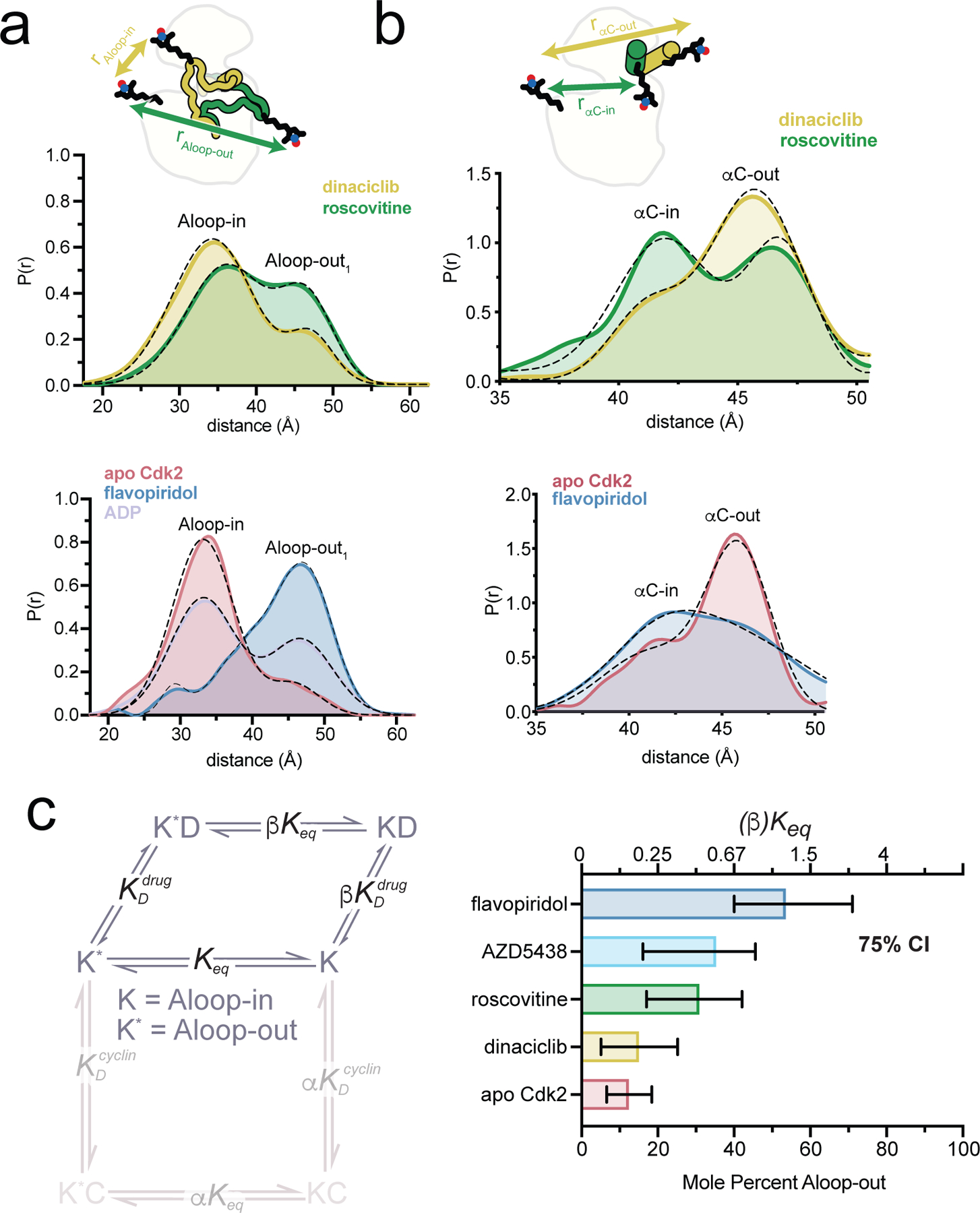
Figure 1:DEER analysis of the Cdk2 A-loop and αC-helix reveals inhibitor-dependent conformational states and supports an allosteric two-state model of Cdk2 activation and inhibitor binding.
Image source: Majumdar et al., Nat. Chem. Biol. (2021), 17, 456–464
Case study 2: Cyclic nucleotide-gated (CNG) ion channels
Cyclic nucleotide-gated (CNG) ion channels play an important role in vertebrate vision and olfaction by converting sensory stimuli into electrical signals. These channels are activated when cyclic nucleotides, such as cAMP or cGMP, bind directly to their intracellular regulatory domains, triggering the opening of the ion-conducting pore. However, the allosteric mechanism by which this occurs is incompletely understood.
The researchers in this case study used double electron–electron resonance (DEER) spectroscopy to examine conformational changes in SthK, a bacterial CNG channel from Spirochaeta thermophila. Spin labels were strategically introduced into the C-linker region, which is known to mediate communication between the cyclic nucleotide-binding domain and the channel gate. The results showed that binding of the full agonist cAMP induced an outward displacement of the B′-helix relative to the channel’s central axis, whereas the partial agonist cGMP failed to produce the same effect. This agonist-dependent structural change was observed in both detergent-solubilized channels and channels incorporated into lipid nanodiscs.
Structural models generated using DEER-derived distance constraints further indicated that channel activation is associated with an upward movement of the cytoplasmic domain and the establishment of specific interactions between the C-linker and transmembrane regions.These findings identify a previously unrecognized conformational transition in CNG channels and provide valuable insight into the structural mechanisms underlying allosteric gating.
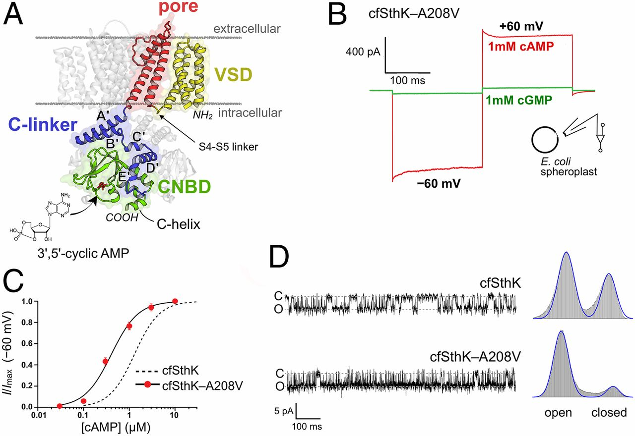
Figure 2. Structure and function of the bacterial CNG channel SthK-A208V. (A) Structure of SthK showing its major domains and the cAMP-binding site. (B) cAMP strongly activates the channel, whereas cGMP produces minimal activation. (C) The A208V mutation increases cAMP sensitivity. (D) Single-channel recordings show a higher open probability for SthK-A208V in the presence of cAMP.
Image source: Evans et al., PNAS 117, 10839–10847 (2020). https://doi.org/10.1073/pnas.1916375117
The Future of Allosteric Drug Discovery
Our understanding of allostery has moved from a largely static view of protein structure toward one in which conformational dynamics and population shifts are central to regulation. This change in perspective is also influencing how we approach allosteric drug discovery. The important question is no longer simply where a ligand binds, but how binding changes the ensemble of states available to the protein and how those changes relate to function. Addressing this question requires different experimental approaches to be used together. EPR, and particularly DEER, is valuable because it provides a direct measure of changes in interspin distances and conformational populations. When combined with structural, biochemical, and computational studies, these measurements can help distinguish ligand-induced conformational changes from pre-existing states and provide a stronger basis for interpreting mechanism of action.
For drug discovery, the key value of this approach is the ability to connect ligand binding with changes in protein conformation and function. Defining how different compounds reshape a target's conformational landscape can strengthen mechanism-of-action studies, improve compound characterization, and provide useful guidance during lead optimization.