Electron paramagnetic resonance spectroscopy is a powerful way to study materials, molecules, and systems with unpaired electrons. It is often called EPR, ESR, electron spin resonance spectroscopy, or spin resonance spectroscopy. The method shows how electron spins respond in a magnetic field, and it helps researchers read structure, motion, bonding, and chemical setting from that response. This guide explains the core ideas behind electron paramagnetic resonance spectroscopy, how EPR spectra form, and why the method matters in chemistry, materials science, biology, and physics.
What is electron spin resonance?
Electron spin resonance is a spectroscopic method that detects changes between spin states of unpaired electrons. In day-to-day use, electron spin resonance, ESR, and electron paramagnetic resonance are often used side by side, with EPR being the most common term in current papers. If you ask what electron spin resonance is, the short answer is simple: it is a way to see how an unpaired electron takes in energy in a magnetic field when microwave energy is added.
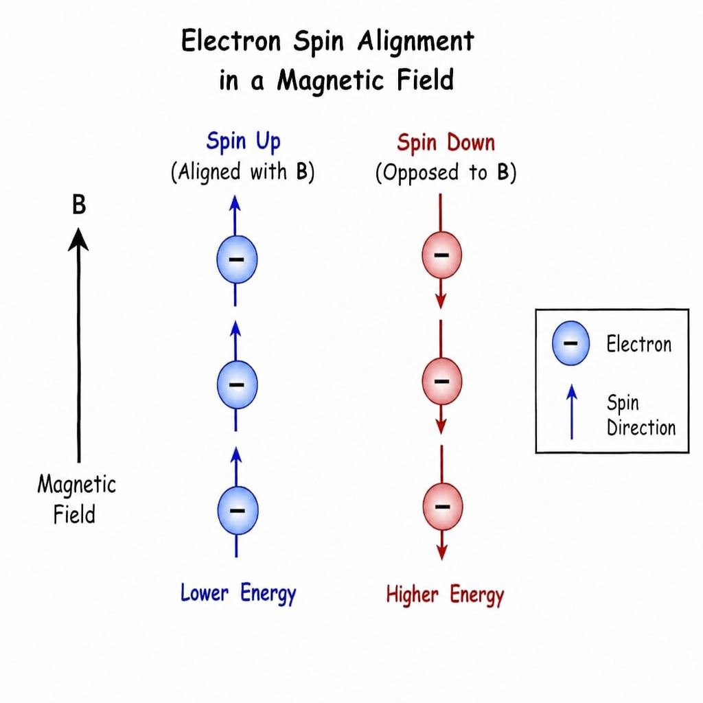
The method rests on a basic property of electrons called spin. When an electron is unpaired, its spin creates a tiny magnetic moment. In an outside magnetic field, that moment can sit in different energy levels. When the microwave energy matches the gap between those levels, resonance happens, and the instrument records a signal.
That signal becomes part of an EPR spectrum. By reading EPR spectra or ESR spectra, scientists can learn about the local setting around the unpaired electron. This can include molecular shape, nearby nuclei, metal centers, defects in solids, or the motion of radicals in a sample.
The basic idea behind spin resonance spectroscopy
Spin resonance spectroscopy works by joining a magnetic field, microwave energy, and the quantum behavior of spin. The sample sits in a resonator inside a magnetic field. As the field is swept or tuned, the instrument watches how strongly the sample absorbs microwave energy.
When absorption occurs, the system has reached electron resonance. The field position, line shape, and splitting pattern all tell us something about the unpaired electron and its setting. This is why electron resonance spectroscopy is useful not only for detecting radicals, but also for studying transition metal ions, defects, catalysts, polymers, semiconductors, and living systems with spin-active centers.
A normal EPR experiment does more than answer whether unpaired electrons are present. It can also show how those electrons interact with nearby atoms, how far they move, and how their chemical setting changes during a reaction or process.
EPR and ESR terms in context
The language around this method can feel messy because several names overlap. Electron paramagnetic resonance spectroscopy, EPR spectroscopy, ESR spectroscopy, and electron spin resonance spectroscopy may all refer to the same family of methods. Some writers even use the mixed label esr epr spectroscopy. Older papers may use electron spin resonance esr spectroscopy as a long form. The term choice often depends on field, habit, or journal style.
ESR points to electron spin resonance and the spin jump itself. EPR points to the wider spin behavior that lets unpaired electrons respond to a magnetic field. In many labs, electron paramagnetic resonance and ESR are used as a matched pair of terms for clarity, especially when talking with people who know one name better than the other.
A useful way to keep the terms straight is this: ESR describes the resonance of electron spin, while EPR describes resonance from spin-active species. In practice, both words point to the same core measurement. Some authors also use electron paramagnetic spectroscopy or electron resonance for the same broad idea.
How does EPR spectroscopy produce useful spectra?
EPR spectroscopy measures microwave absorption from unpaired electrons. This absorption occurs as their spin states change in a magnetic field. The process produces useful spectra. The pattern is not random. It reflects the magnetic, electronic, and structural setting around the unpaired electron. A well-read spectrum can help identify a species, compare samples, or track changes as conditions shift.
Several features make EPR spectra useful:
- Resonance position: The field value where absorption appears can suggest the type of electron setting and whether the electron sits in an organic radical, a metal complex, or a solid defect.
- Signal strength: Stronger signals can mean more spin-active species, although sample state and instrument settings also matter.
- Line shape: Narrow, broad, even, or distorted lines may show motion, interactions, concentration effects, or mixed environments.
- Hyperfine splitting: Interactions between the unpaired electron and nearby nuclei can create several lines, which gives clues about molecular shape.
- Angle effects: In solids, crystals, or fixed samples, spectra may change with angle, revealing directional electronic behavior.
This mix of features makes electron paramagnetic spectroscopy especially useful when other lab tools give only indirect clues. For example, a color change in a reaction may suggest a radical step, but electron paramagnetic resonance spectroscopy can more directly test whether an unpaired electron is present.
Main parts of an EPR experiment
Although instruments vary, most EPR systems use the same basic parts. Knowing these parts helps explain why the method is sensitive to both the sample and the setup.
Magnetic field system
The magnetic field separates the energy levels linked to unpaired electron spin. By changing the field strength, the instrument finds the point where microwave energy matches the spin jump. Field stability and calibration matter because even small shifts in resonance position can carry useful information.
Microwave source and resonator
Microwave energy supplies the needed push for spin changes. The resonator holds the sample in a place where microwave interaction is strong. Sample size, shape, placement, and solvent choice can all affect signal quality, so sample prep matters.
Detector and signal processing
The detector records changes in microwave absorption, and the output is often a derivative-shaped spectrum rather than a plain absorption peak. Signal processing can improve visibility, but it must be used with care. Too much processing can make spectra look neat while hiding useful detail.
Sample setting
Temperature, atmosphere, light, and sample phase can all change the observed spectrum. Some radicals are short-lived, some metal sites change oxidation state, and some materials behave very differently at low temperature. Good test design keeps these points in mind.
Common uses of electron paramagnetic resonance
Electron paramagnetic resonance has broad value because unpaired electrons appear in many science and industry settings. The method is not limited to one sample type or one research question.
Common uses include:
- Free radical detection
EPR can find and study radicals formed during oxidation, light-driven reactions, polymer breakdown, burning, or biochemistry. - Transition metal analysis
Many metal ions have unpaired electrons. EPR can give insight into coordination setting, oxidation state, and ligand interactions. - Catalyst study
Catalysts often pass through spin-active middle steps. EPR can help track these states and map reaction paths. - Materials and defect studies
Defects in crystals, glasses, chips, and nano materials may contain unpaired electrons. EPR can probe these sites even when bulk methods miss them. - Biological and biochemical research
Metal proteins, enzymes, spin-labeled biomolecules, and radical processes can be studied with EPR when the test setup is right. - Radiation and dose studies
Some exposed samples form stable spin-active centers. EPR can detect these centers and support work on radiation effects.
Reading EPR spectra with care
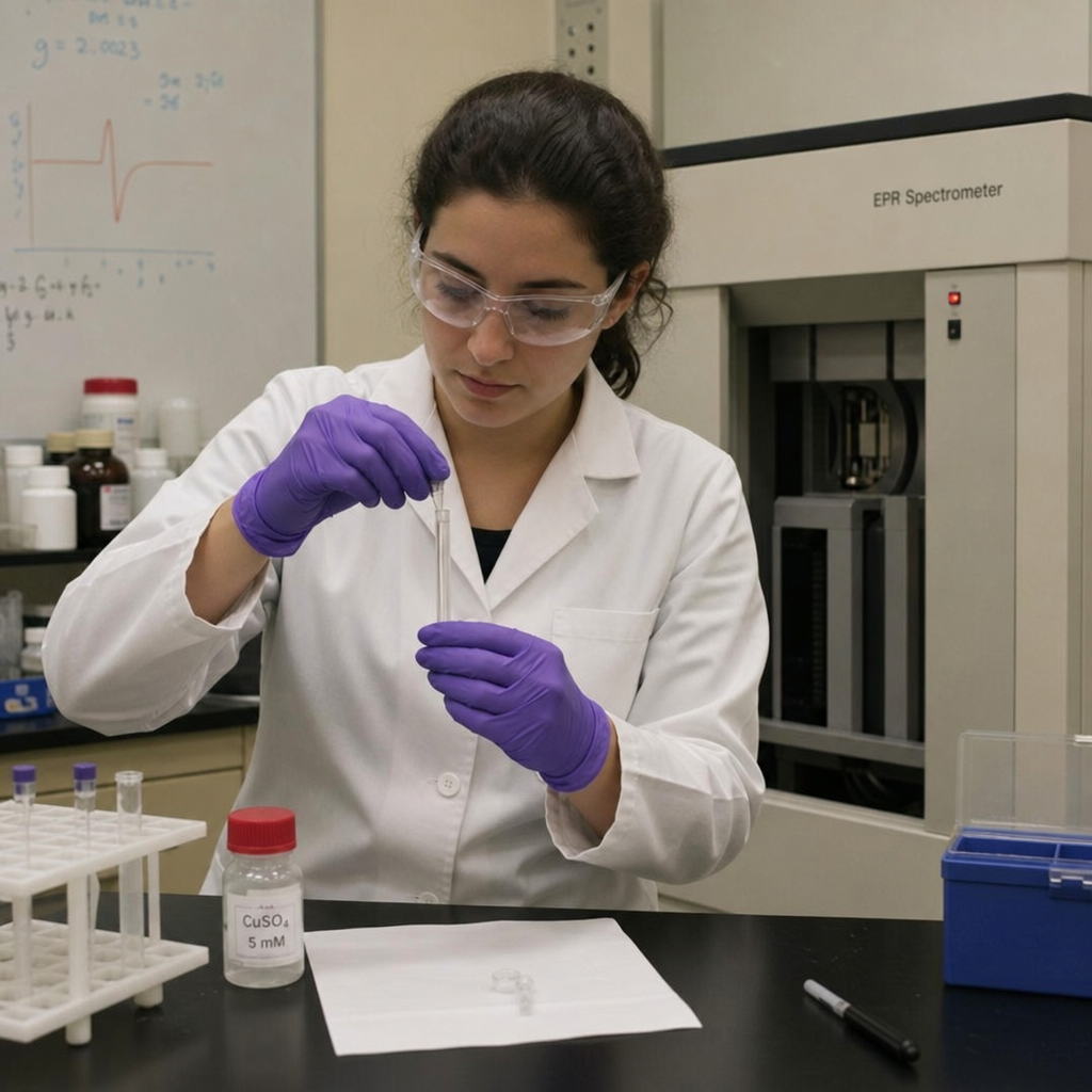
At first glance, EPR spectra may look like a set of peaks and dips, but each feature can hold layered meaning. A single clean signal may point to one main spin-active species, while complex splitting can show links with several nuclei. Broad signals can point to strong interactions, short relaxation times, or mixed environments. Still, the reading step needs care. Similar-looking spectra can come from different systems, and small changes in instrument settings may change the look. Researchers usually compare spectra with reference data, simulations, control samples, and other methods before they draw a firm conclusion.
A practical reading workflow might include:
- Confirm that the signal comes from the sample, not the tube, solvent, cavity, or stray material.
- Record controls, such as a blank or untreated sample.
- Check whether signal strength changes with concentration, temperature, light, or reaction time.
- Look at splitting patterns and line widths, not just peak position.
- Use simulation or reference data when assigning complex spectra.
- Read EPR results with chemical context and other lab data.
This disciplined approach helps prevent overstatement. EPR is highly informative, but it works best as part of a wider evidence set.
Why EPR stands out among spectroscopy methods
EPR is valuable because it is selective for unpaired electrons, while many other spectroscopy methods respond to broader molecular or structural features. That focus makes it especially useful when the species of interest is a radical, a spin-active ion, or a defect site in a more complex material. Instead of averaging only the bulk mix, EPR can focus on electronically active centers.
Compared with UV-visible, infrared, or nuclear magnetic resonance spectroscopy, EPR answers a different kind of question. It is less about all bonds or all nuclei in a sample and more about where unpaired electron density sits and how it interacts with its surroundings. This makes it helpful in systems where spin-active species control reaction, performance, breakdown, or biological function.
The method also fits well with imaging, separation, electrochemistry, and computer modeling. For example, a reaction study may use separation to track products and EPR spectroscopy to watch a radical step. Together, the methods can give a fuller picture than either one alone.
Practical things to check before using EPR
Consider if the sample has unpaired electrons before picking electron resonance or electron paramagnetic resonance spectroscopy. Materials with all paired electrons usually do not give a standard EPR signal. If the target species is short-lived, unstable, or present at low level, sample handling becomes very important.
Researchers should also think about whether room-temperature work is enough or whether low-temperature work may improve signal stability and resolution. Solvent, oxygen, concentration, and sample shape can all affect the result. In solid samples, angle and mixed structure may make spectra harder to read, but they can also reveal useful detail.
A simple pre-test checklist includes:
- Is the species expected to be spin-active?
- Could oxygen, moisture, or light change the sample?
- Is a liquid, frozen solution, powder, film, or crystal the best choice?
- Are controls available for comparison?
- Will the expected signal be broad, weak, or overlapped?
- Is extra analysis needed to support the reading?
Thinking through these questions early can save time and make the final data more reliable.
The larger role of EPR in modern analysis
As materials, catalysts, energy systems, and biological models get more complex, tools that can isolate key electronic features become more useful. Electron paramagnetic resonance remains important because it gives researchers a direct view of unpaired electrons, often the same species that drive reaction, failure, or function.
The method is also flexible. Forms of EPR can look at distance, motion, pulsed tests, spin labeling, and advanced materials study. Even when a project starts with a simple question such as 'is there a radical here?', the method can lead to deeper insight about mechanism and structure.
For students and new users, the best place to start is the link between spin, magnetic field, microwave absorption, and spectrum shape. Once that base is clear, the many names ; electron resonance, ESR spectroscopy, EPR spectroscopy, and electron paramagnetic resonance become less daunting.

Electron paramagnetic resonance spectroscopy is a very useful form of spin resonance spectroscopy for studying unpaired electrons. Whether called EPR, ESR, electron spin resonance spectroscopy, or electron resonance spectroscopy, the method turns magnetic interaction into readable spectral patterns. For any sample where radicals, transition metals, defects, or other spin-active centers matter, EPR can give evidence that other methods may miss. Its real strength comes from careful test design, good controls, and reading the spectrum in the full chemical or material context.