AlphaFold2 has transformed how researchers predict protein structure, but understanding how proteins move between conformations requires experimental information. Electron Paramagnetic Resonance (EPR) spectroscopy, particularly Double Electron-Electron Resonance (DEER) spectroscopy provides a way to measure those conformational changes and complement AlphaFold predictions.
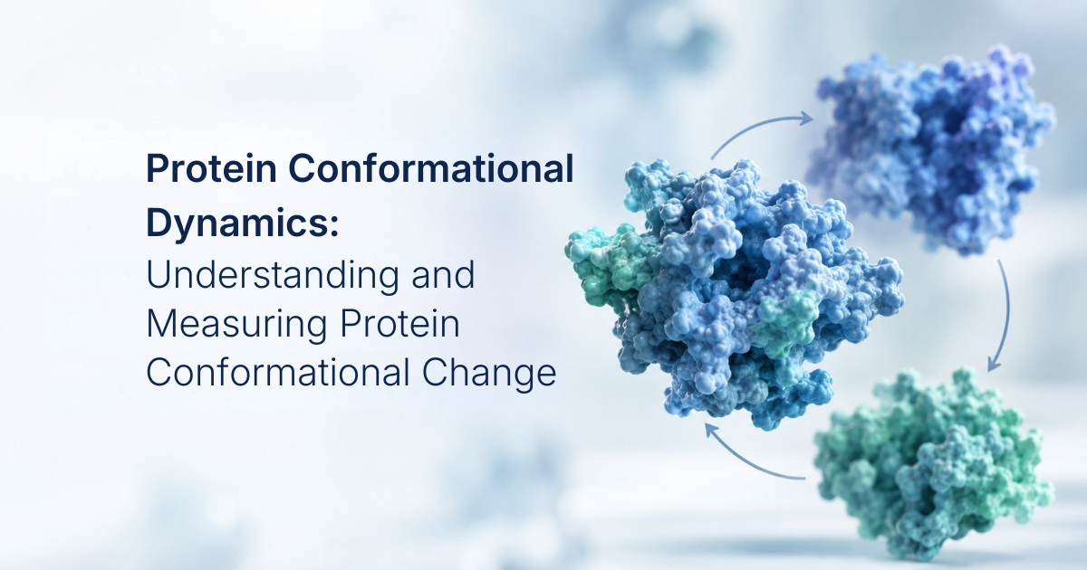
Few tools have changed structural biology as quickly as AlphaFold2. Within a few years of its release, AlphaFold2 had become an important starting point for structural studies of many proteins. However, transporters, receptors, channels and many enzymes function through conformational changes, and ligand binding can shift proteins between different structural states. A single predicted model, however confident, represents one structural state.
This is where Electron Paramagnetic Resonance (EPR) spectroscopy comes in. In this post I will be walking through why AlphaFold and EPR spectroscopy, in particular the pulsed distance technique known as Double Electron-Electron Resonance (DEER) spectroscopy, make such natural partners, following the three threads in the title: structure, dynamics, and function.
Protein Structure: what AlphaFold gives us, and what it doesn't
I'll begin with what AlphaFold2 was built to do: predict a three-dimensional protein structure from its amino acid sequence and related sequence information. Wu and colleagues describe AlphaFold2 as predominantly assigning a single conformation to a given input sequence. AlphaFold2's predictions also depend on information contained in the multiple sequence alignment (MSA), while standard AlphaFold2 does not directly describe the full conformational ensemble of a protein. AlphaFold2 also does not directly predict minor alternative conformations or the effects of mutations, post-translational modifications, or ligand binding.
The community has responded with several approaches for sampling alternative conformations. By using shallow or subsampled MSAs, or by systematically modifying the MSA as in the SPEACH_AF method, researchers have generated models spanning alternative states, including inward-facing and outward-facing states of membrane proteins. These ensembles are useful structural hypotheses, but they are predictions. They do not by themselves establish which states are populated experimentally, their relative populations, or how those populations change under different biochemical conditions.
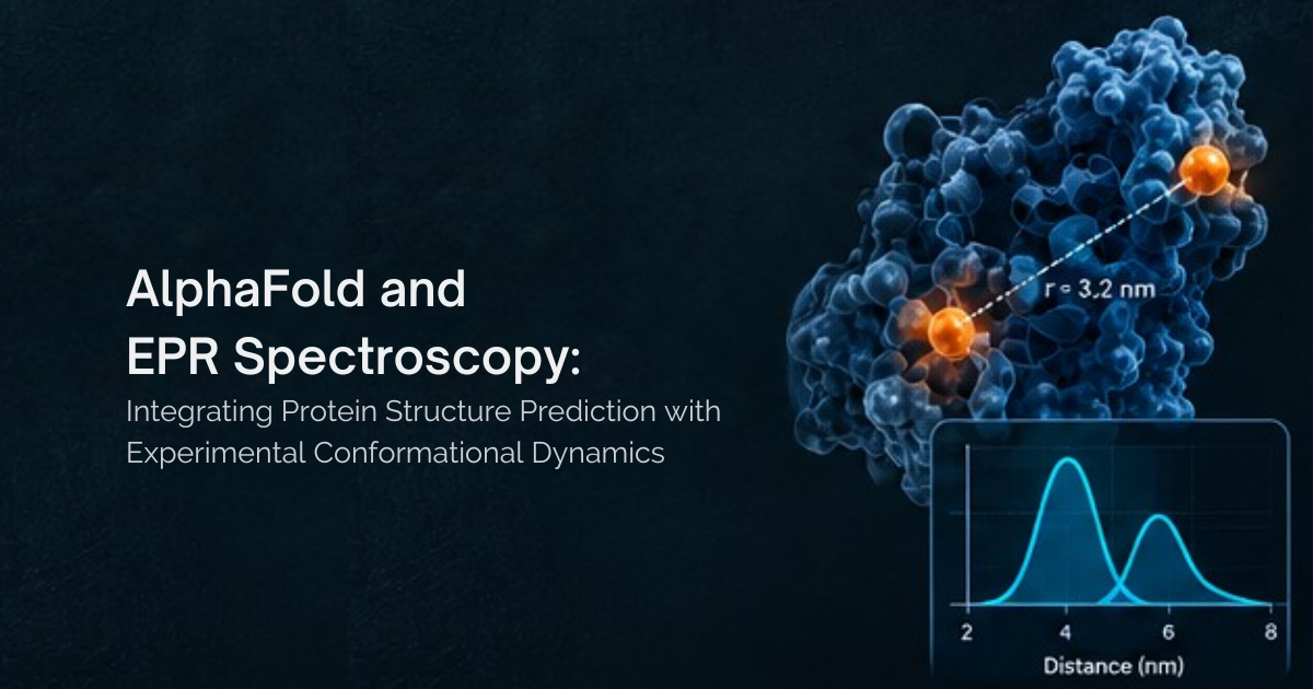
Answering those questions requires experimental measurements, and EPR spectroscopy is particularly well suited to studying protein structure and conformational dynamics. Most proteins do not contain the unpaired electrons required for conventional EPR measurements, so paramagnetic centers can be introduced through site-directed spin labeling (SDSL), pioneered by Wayne Hubbell and colleagues. In a common SDSL strategy, cysteine residues are introduced at selected sites and reacted with a nitroxide spin label such as MTSSL. With two labels in place, DEER spectroscopy measures the magnetic dipolar interaction between the spin labels and provides a distance distribution. DEER commonly accesses distances of approximately 15–80 Å. The fact that DEER provides a distribution rather than simply a single mean distance makes it particularly useful for investigating conformational heterogeneity.
Protein Dynamics: from one model to an ensemble
When different protein conformations produce distinct distance populations, DEER can reveal multiple components in the measured distance distribution. With appropriate structural modeling and analysis, the relative amplitudes of these components can provide information about the populations of the corresponding conformational states. Because experimental conditions can be varied, researchers can add a substrate, inhibitor, ion, or nucleotide, change the pH, or examine a membrane protein in different environments such as detergent micelles and lipid nanodiscs, and measure how the distance distribution responds.
One limitation is that each spin-label pair probes the distance between one selected pair of sites, so multiple label pairs are often needed to build a broader structural picture. Connecting those distance measurements to specific three-dimensional conformations therefore benefits from a structural framework and appropriate modeling. AlphaFold2-based ensemble-generation methods can provide additional structural hypotheses that help place these experimental distance measurements into a three-dimensional context.
A nice illustration comes from the study of GadC, a pH-dependent amino acid/polyamine/organocation (APC) antiporter. GadC contributes to bacterial acid resistance by exchanging extracellular glutamate for intracellular γ-aminobutyric acid (GABA). Its experimentally determined structure represented an inward-facing state, but this structure alone could not establish how the transporter moves between inward- and outward-facing conformations during its transport cycle.
Del Alamo and colleagues combined DEER spectroscopy with an AlphaFold2-based approach that generated structural models representing multiple conformations of GadC. The resulting models were used to guide the selection of experimental labeling sites and to facilitate interpretation of the DEER measurements. DEER experiments performed in lipid bilayers then monitored how the transporter responded to acidification and substrate binding. The measurements revealed acid-induced structural rearrangements involving the C terminus and transmembrane helices and provided evidence for transitions between inward- and outward-facing states. Glutamate produced a different effect, modulating the dynamics of an extracellular gate without substantially shifting the overall inward/outward equilibrium.
A second example, MhsT, is a bacterial homolog/ortholog of the neurotransmitter:sodium symporter (NSS) family. NSS transport substrates across membranes through coordinated conformational changes, but the intermediate states involved in this process are difficult to capture using static structures alone. Schwartz and colleagues used SPEACH_AF to generate clusters of MhsT structures spanning the transition between inward- and outward-facing conformations. DEER spectroscopy was then used to monitor movements of multiple structural motifs under different ligand conditions and in two different membrane environments: detergent micelles and lipid nanodiscs. The experiments showed that ligand binding altered specific structural movements and that the surrounding membrane environment influenced the energetics of those conformational changes. The finding I find most striking is that running the same measurements in detergent micelles and in lipid nanodiscs revealed a profound effect of the environment on the energetics of those motions. For anyone who screens or solves structures in detergent, that is worth pausing on.
Put together, these studies settle into a workflow that I think is worth sketching out. AlphaFold-based ensemble methods generate structural hypotheses, and in doing so also tells us, before any protein is labeled, which residue pairs will best tell those states apart. DEER spectroscopy then provides experimental evidence for which conformational states are populated under the conditions tested.
The most recent turn of this cycle closes the loop entirely. In 2025, Wu and colleagues introduced DEERFold, a modified version of AlphaFold2 that incorporates experimental DEER distance distributions into the protein structure prediction process. The authors investigated whether spin-label distances derived from DEER data could serve as direct input to guide AlphaFold2 predictions.
The authors' goal was to develop and benchmark a method that could guide AlphaFold2 toward multiple protein conformations consistent with experimental data. By incorporating DEER-derived distance distributions into the prediction process, DEERFold can guide predictions toward experimentally characterized target conformations.
The authors first tested DEERFold using two membrane transporters: LmrP, a proton-coupled antiporter of the major facilitator superfamily (MFS), and PfMATE, a proton-coupled multidrug transporter from the multidrug and toxic compound extrusion (MATE) superfamily. LmrP's proton-powered conformational cycle had previously been investigated using DEER in detergent micelles and nanodiscs, providing experimental distance distributions for benchmarking DEERFold. For PfMATE, the authors likewise used DEER-derived distance information to test whether the method could guide predictions between different conformational states.
For both transporters, the unconstrained predictions favored the outward-facing conformation, whereas incorporating DEER-derived distance information could guide the predictions toward the inward-facing target conformation. For LmrP, the authors also tested extremely sparse sets of simulated distance constraints. With only two optimized spin-label distance constraints, 58 of 100 models approached the target state with Root Mean Square Deviation (RMSD) values below 3 Å.
Wu and colleagues then evaluated DEERFold across a broader collection of soluble and membrane proteins and applied the method to the multidomain ABC transporter P-glycoprotein (Pgp/ABCB1). The authors found that a limited number of optimized spin-label distance constraints could guide predictions toward target conformations. Their analyses also showed that the particular distance constraints selected mattered: constraint sets containing key structural information were more effective at driving the models toward the target conformation.
Function: why this matters for drug discovery
For those of us designing molecules, the value of combining AlphaFold with DEER is that it can provide experimental evidence for which conformational states are populated under defined conditions. This creates the possibility of using experimentally supported conformations as additional structural hypotheses for structure-based drug discovery. Ensemble-based approaches are already used in drug discovery to explore multiple protein conformations, including for allosteric and cryptic-site discovery.
DEER measurements can also reveal how ligand binding or changes in conditions affect protein conformations. In GadC, for example, acidification shifted the protein between inward- and outward-facing conformations, while glutamate produced a more localized effect at an extracellular gate without substantially shifting the overall inward/outward equilibrium. This illustrates how DEER can distinguish between broader conformational rearrangements and more localized structural responses.
The MhsT study provides another important example. Comparative DEER measurements in detergent micelles and lipid nanodiscs revealed a profound effect of the surrounding environment on the energetics of conformational changes. For membrane-protein drug discovery, this suggests that comparing conformational behavior under different sample conditions can provide useful information before selecting an experimental system for downstream studies.
There is also a data story here for AI-driven discovery. Experimentally determined structures generally provide snapshots of particular conformational states, whereas DEER distance distributions can provide information about structural heterogeneity under defined conditions. DEERFold demonstrates that experimental DEER distance information can be incorporated directly into an AlphaFold2-based prediction process to guide predictions toward experimentally supported conformations.
References
- Wu T, Stein RA, Kao T-Y, Brown B, Mchaourab HS. Modeling protein conformational ensembles by guiding AlphaFold2 with Double Electron Electron Resonance (DEER) distance distributions. Nat Commun. 2025;16:7107.
- Schwartz AC, Stein RA, Gil-Iturbe E, Quick M, Mchaourab HS. Alternating access of a bacterial homolog of neurotransmitter:sodium symporters determined from AlphaFold2 ensembles and DEER spectroscopy. Proc Natl Acad Sci USA. 2024;121(40):e2406063121.
- del Alamo D, DeSousa L, Nair RM, Rahman S, Meiler J, Mchaourab HS. Integrated AlphaFold2 and DEER investigation of the conformational dynamics of a pH-dependent APC antiporter. Proc Natl Acad Sci USA. 2022;119(34):e2206129119.
- Laurents DV. AlphaFold 2 and NMR spectroscopy: partners to understand protein structure, dynamics and function. Front Mol Biosci. 2022;9:906437.

There is a great deal more to say on this topic, from in-cell DEER to the steady stream of new AlphaFold sampling methods. My hope is that this post serves as a starting point. AlphaFold has made structural hypotheses abundant and cheap; for dynamic targets, the harder question is now which of those hypotheses describe the protein a drug will actually meet. Paired with EPR spectroscopy, AlphaFold moves us from a single picture of structure toward a measured view of dynamics, and from there toward a much clearer understanding of function. For anyone building a programme on a predicted structure, it is worth asking which conformation the chemistry is meant to bind, whether there is evidence that conformation is populated, and whether anyone has measured how the compound shifts the balance. Till next time!