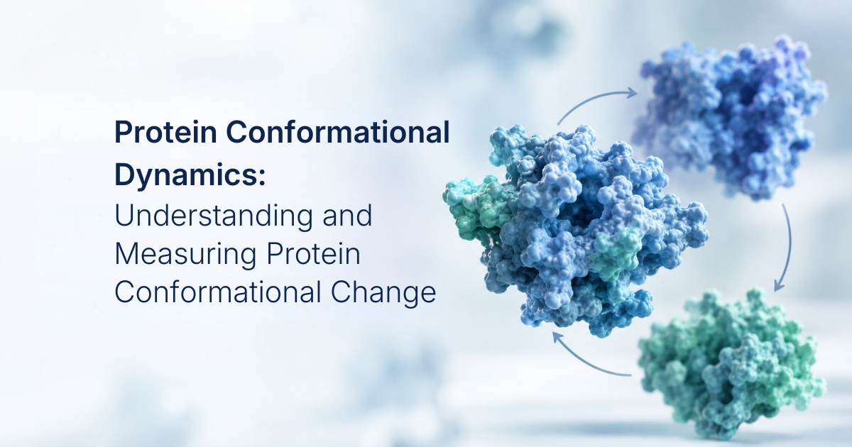
Protein conformational dynamics describe the ability of proteins to shift between different three-dimensional structures in response to factors such as ligand binding, environmental conditions, post-translational modifications, and interactions with other molecules. These transitions generally occur without breaking covalent bonds and instead involve changes in noncovalent interactions, including hydrogen bonding, electrostatic interactions, hydrophobic effects, and van der Waals forces.
Although these structural rearrangements can be relatively small, they can have substantial functional consequences. A change in conformation may alter the accessibility of a binding site, reposition catalytic residues, modify interactions between protein domains, or shift the equilibrium between functional states. Protein function therefore often depends on the ability of the molecule to sample and transition between multiple conformational states.
Why Do Protein Conformational Dynamics Matter?
Protein structure and dynamics are closely connected to biological function. Conformational transitions contribute to processes such as enzyme catalysis, allosteric regulation, signal transduction, molecular transport, and protein–protein interactions. Understanding these structural changes is therefore important in structural biology, therapeutic development, and biopharmaceutical research. A key challenge is that proteins can exist as ensembles of interconverting conformational states and the relative populations of these states can change with ligand binding, mutations, environmental conditions, or interactions with other molecules. Some states may also be transient or sparsely populated, making them difficult to detect using methods that primarily provide structural snapshots.
What Causes Protein Conformational Changes?
Ligand Binding
Ligand binding is a common driver of protein conformational change. Substrates, ions, peptides, drugs, and other molecules can alter the equilibrium between different structural states. In enzymes, substrate binding can promote structural rearrangements that position catalytic residues for productive chemistry. In receptors and transport proteins, ligand binding can shift the protein toward states associated with signaling or molecular transport.
Allosteric Regulation
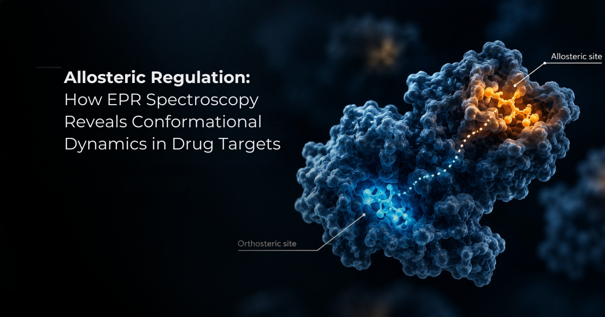
Allosteric regulation occurs when binding at one region of a protein influences the structure or activity of another site. Rather than directly occupying the catalytic or primary ligand-binding site, an allosteric molecule can shift the equilibrium between different conformational states. This provides a mechanism for coupling molecular recognition at one location to functional changes elsewhere in the protein and is particularly important in enzymes and signaling proteins.
Post-Translational Modifications
Post-translational modifications can alter protein conformational behavior by changing the chemical properties of specific residues. Modifications such as phosphorylation, acetylation, methylation, and ubiquitination can affect intramolecular and intermolecular interactions and consequently influence protein activity, localization, molecular interactions, or degradation. In signaling pathways, these modifications can shift the balance between different functional states.
Environmental Conditions
Protein conformations are also sensitive to environmental factors, including pH, temperature, ionic strength, and solvent composition. Changes in pH, for example, can alter the protonation state of amino acid side chains and affect electrostatic interactions. Temperature changes can influence molecular fluctuations and the stability of different conformational states. Depending on the conditions, these effects may produce subtle structural rearrangements or, under more extreme conditions, partial or complete unfolding.
Mechanical Forces
Mechanical forces can drive conformational transitions in proteins involved in molecular movement and force generation. Myosin and kinesin, for example, undergo coordinated structural changes during ATP binding, hydrolysis, and product release. These transitions regulate their interactions with actin or microtubules and enable chemical energy to be coupled to directional mechanical movement.
Examples of Protein Conformational Changes
Protein conformational dynamics can be observed across many biological systems. Several well-characterized examples demonstrate how structural transitions are linked to molecular function.
Enzymes
Many enzymes undergo conformational rearrangements during substrate binding and catalysis. These changes can improve substrate positioning and alter the configuration of catalytic residues. In some enzymes, conformational dynamics help populate catalytically competent states during the reaction cycle.
Hemoglobin
Hemoglobin is a classic example of conformational coupling between ligand binding and protein function. Oxygen binding shifts the equilibrium toward the higher-affinity R state relative to the lower-affinity T state. Because the four subunits are structurally coupled, ligand binding at one site influences the affinity of other sites, producing cooperative oxygen binding.
G-Protein-Coupled Receptors
G-protein-coupled receptors (GPCRs) use conformational changes to translate extracellular ligand binding into intracellular signaling. Ligand binding can shift a receptor among different conformational states, influencing interactions with signaling partners such as heterotrimeric G proteins and arrestins. Different ligands can preferentially stabilize different receptor states, contributing to differences in downstream signaling.
Membrane Proteins
Conformational dynamics are particularly important for membrane proteins, including receptors, ion channels, and transporters. Structural transitions can regulate processes such as channel opening, substrate transport, and signal transmission. Because these proteins operate within complex membrane environments, characterizing their conformational states under relevant conditions can be experimentally challenging.
Why Is Protein Conformational Dynamics Difficult to Measure?
Proteins can occupy multiple conformational states that coexist in equilibrium and interconvert over different timescales. Some states may be highly populated, while others are transient or present only at low abundance. Consequently, a single structural measurement may provide only a partial view of the protein's conformational landscape. Experimental observations can also depend on sample preparation, labeling strategies, temperature, buffer composition, ligand concentration, and other conditions. These factors make it important to distinguish between observing a particular structural state and determining how different states are populated under biologically relevant conditions. For this reason, protein conformational analysis often benefits from combining complementary structural, biophysical, and computational methods.
Methods for Studying Protein Conformational Dynamics
Different techniques provide different types of information about protein structure and dynamics.
X-Ray Crystallography
X-ray crystallography provides high-resolution structural information and can reveal differences between proteins captured under different biochemical conditions. Comparing structures obtained with and without a ligand, for example, can identify local rearrangements or larger domain movements. However, crystallization conditions can influence conformational populations, and transient or highly flexible states may be difficult to characterize.
NMR Spectroscopy
Nuclear magnetic resonance (NMR) spectroscopy is particularly useful for investigating protein structure and dynamics in solution. It can provide information about flexible regions, conformational exchange, and transitions between multiple states. NMR can therefore reveal dynamic behavior that may not be apparent from a single static structure.
Cryo-Electron Microscopy
Cryo-electron microscopy (cryo-EM) has become an important technique for studying large proteins and macromolecular complexes. Modern cryo-EM analysis can identify structural heterogeneity and, in suitable systems, resolve multiple conformational states. However, the structures recovered from a cryo-EM experiment must be interpreted within the context of the experimental conditions and should not automatically be considered a direct representation of conformational populations in solution.
Fluorescence-Based Techniques
Fluorescence methods can monitor structural rearrangements through changes in fluorescent signals. Förster resonance energy transfer (FRET), for example, measures changes in the proximity of fluorescent probes and can therefore report on protein conformational dynamics in vitro and in living cells. However, fluorescent labeling, probe orientation, photophysical effects, and measurement conditions can influence the interpretation of FRET data.
Molecular Dynamics Simulations
Molecular dynamics (MD) simulations provide a computational approach for examining how molecular structures evolve over time. Simulations can identify potential intermediate states, transient conformations, and interactions associated with structural transitions. When combined with experimental measurements, MD simulations can help develop and test models of protein conformational behavior.
EPR Spectroscopy
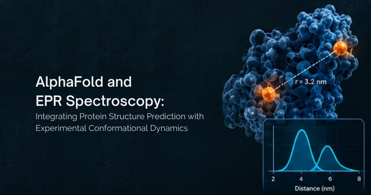
Electron paramagnetic resonance (EPR) spectroscopy provides a complementary approach for studying protein structure and dynamics using paramagnetic centers. Through site-directed spin labeling, paramagnetic probes can be introduced at selected positions within a protein. EPR measurements can then provide information about local mobility, the spin-label environment, and, with appropriate pulsed EPR techniques, distances between labeled sites.This makes EPR useful for investigating conformational changes that may occur without major changes to the overall protein fold. By comparing measurements obtained under different conditions, such as in the presence and absence of a ligand, researchers can examine changes in structural behavior and conformational heterogeneity.
How Does DEER Spectroscopy Measure Protein Conformational Change?
Double electron–electron resonance (DEER), also known as pulsed EPR or PELDOR, is a technique used to measure distances between paramagnetic centers. In protein studies, site-directed spin labeling places paramagnetic probes at selected positions. The dipolar interaction between these spins provides information about the distance separating the labels, typically on the nanometer scale. If two labeled regions move relative to one another during ligand binding or another structural transition, the corresponding DEER distance distribution can change. Comparing these distributions under different biochemical conditions can therefore provide experimental evidence for conformational rearrangement.DEER produces a distribution of distances that can reveal broad or multiple distance populations and provide information about conformational heterogeneity. Measurements from several labeling sites can generate complementary structural restraints that help distinguish between possible conformational models.
Why Do Protein Conformational Dynamics Matter in Drug Discovery?
Protein conformational dynamics are particularly relevant to drug discovery because structural flexibility can influence ligand recognition, binding-site accessibility, and interactions with therapeutic molecules. Different conformational states may expose, close, or reshape binding pockets, while transient structural states can sometimes reveal binding sites that are not apparent in a single static structure. Characterizing these states can contribute to target validation, structure-based drug discovery, and the investigation of protein–ligand interactions. Understanding how a drug candidate affects the conformational equilibrium of its target may also provide information about its mechanism of action and binding behavior.