Techniques like Nuclear Magnetic Resonance (NMR) spectroscopy have long been a cornerstone of structural biology, providing atomic-level insight into protein structure and dynamics in solution. NMR remains one of the most powerful tools for studying folded protein domains, as well as local, site-specific dynamics in partially disordered and flexible regions.
However, as biological systems become larger, more dynamic, or more heterogeneous, challenges begin to emerge. Spectra become increasingly crowded, sensitivity drops, and overlapping signals make interpretation difficult. In many cases, low-population conformations are averaged out and made effectively undetectable. In such cases, how does one access the structural information that falls outside the limits of NMR? The answer to that question usually involves the introduction of a complementary technique to help fill in the gaps. This is where EPR spectroscopy shines.
In this post, we explore how EPR spectroscopy, particularly pulsed dipolar methods, complements NMR to reveal protein flexibility, conformational changes, and ligand-induced dynamics.
What Is EPR Spectroscopy and why is it important for Structural Biology?
Electron Paramagnetic Resonance (EPR) spectroscopy shares the same fundamental underpinnings as NMR. Both techniques apply an external magnetic field to polarize a spin system and drive transitions using electromagnetic radiation. The key difference is that EPR detects electron spins rather than nuclear ones; specifically; unpaired electron spins in the form of stable organic radicals or paramagnetic metal ions.
Most proteins do not naturally contain unpaired electrons. This has two major implications for EPR spectroscopy. The first is that EPR signals typically don’t suffer from background signals, making measurement and data interpretation straightforward. The next is that most proteins and biomolecules are not natively detectable to EPR. To make a protein EPR-detectable, one must introduce a stable paramagnetic species deliberately and strategically, through engineered sites on the protein. The technique for doing this is called site-directed spin labeling, or SDSL. Using pulsed EPR techniques such as DEER (Double Electron–Electron Resonance), sometimes known as PELDOR (Pulsed Electron Electron Double Resonance), it becomes possible to measure distances between two labeled sites. Notably, these techniques do not return just a mean distance, but provide a probability distribution of distance which directly reflect the underlying conformational ensemble of the protein.
Learn about spin labels here: Spin Class: EPR’s Long Distance Relationships
Pulsed Dipolar Spectroscopy (DEER/PELDOR)
In the DEER experiment, two spin labels on a protein experience a dipolar interaction whose strength depends on the distance between them. DEER isolates that interaction through a timed sequence of nanosecond-scale microwave pulses applied to the spin labeled protein sample. The resulting signal encodes the distance between them, which can be extracted as a distribution. These distributions provide structural and dynamic insights:
- Narrow distributions → rigid, well-defined conformations
- Broad distributions → flexible or disordered regions
- Multiple peaks → distinct conformational states coexisting in equilibrium
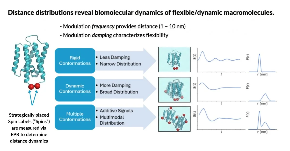
The distribution itself is a direct readout of how the protein conforms under the experimental conditions. In other words, EPR does not just tell you
what structure exists, it tells you how many conformation states exist, and how they are distributed in solution because the experiment captures all conformations present at the moment of flash freezing. Thus, multiple states can be observed simultaneously in a single measurement. This makes DEER a direct and powerful experimental tool for studying protein conformational dynamics.
How EPR Spectroscopy Adds Complementary Insights to NMR
1. Long-Range Distance Constraints Beyond NOE Range and large biomolecules
NMR nuclear Overhauser effect (NOE)- based restraints operate at short range, typically under ~0.6 nm. While paramagnetic relaxation enhancement (PRE) can extend this, it has limitations. In contrast, DEER operates in the ~2–8 nm range, providing long-range distance restraints that complement NMR’s short-range data. Additionally, determining protein structures using NMR for proteins larger than 60-100 kDa often proves prohibitively complex, whereas EPR is not limited by protein size. This makes EPR especially useful for domain movements, multi-domain proteins, and large complexes.
2. Intrinsically Disordered Proteins and Regions (IDPs and IDRs)
Broad linewidths, spectral overlap, and extreme conformational heterogeneity make assignment and structural interpretation genuinely difficult for highly disordered systems through NMR. However, DEER reports distance distributions directly, providing a readout of conformational heterogeneity. Changes in the distribution directly reflect compaction or disorder-to-order transitions under conditions such as ligand binding or partner interaction.
3. Multiple Conformational States Captured Simultaneously
One of DEER’s most distinct capabilities is capturing multiple coexisting conformational states in a single measurement. As an ensemble measurement technique, DEER detects all species that are present at the time of flash freezing. If your protein has multiple states that exist in equilibrium, they can appear as separate peaks in the distance distribution, with relative areas proportional to their populations. This makes EPR a powerful tool for studying allosteric regulation, conformational switching, and ligand-induced structural changes.
4. Membrane Proteins
Solution NMR is limited by protein size and solid-state NMR is powerful but technically demanding. Similarly, DEER is a powerful but specialized technique; however, it has become a primary tool for studying membrane protein conformational dynamics because it has no size restriction and has been widely applied to transporters, ion channels, GPCRs, and other membrane proteins.
5. Metalloproteins
For researchers working with metalloproteins, EPR offers something unique: the metal itself can serve as the spin probe. If your protein naturally contains a paramagnetic metal ion such as copper, iron, manganese or cobalt, they already carry unpaired electrons that EPR detects directly without spin labeling. This gives you direct access to the metal binding pocket geometry, coordination environment, and local electronic structure.
Why NMR Users Should Embrace EPR for Protein Dynamics
- It’s complementary: EPR adds what NMR can’t see, without replacing the residue-level insight you already trust.
- It’s ensemble-friendly: Flexible regions, disordered domains, and transient interactions are all captured naturally.
- It’s scalable: Large proteins, complexes, and membrane proteins that challenge NMR can be probed efficiently.
- It accelerates insight: Low-population states, ligand effects, and conformational heterogeneity can be rapidly characterized with a few strategic spin labels.
FATHOM EPR + NMR: A Practical Complement for Modern Structural Biology
FATHOM removes the traditional barriers to EPR allowing NMR and structural biology labs to access complementary distance and ensemble information without requiring deep EPR expertise. This is accomplished by simplifying and automating the workflow:
- Automated experiment setup, tune and calibration
- Integrated data acquisition and analysis
- Automated reporting and distance measurements
- Minimal sample requirements (~3.5µL)
- Ability to run multiple samples unattended
Historically, EPR analysis has required specialized expertise but FATHOM EPR changes that.
Learn more about FATHOM EPR here: FATHOM EPR Spectrometer | High Q Technologies
Interested in adding EPR spectroscopy to your NMR or structural biology workflow? Contact us to discuss your protein system and how FATHOM can complement your existing research.

In that spirit, what’s most compelling is not just how these approaches extend what we can measure, but how they reshape how we think about structure itself. By capturing distributions and coexisting states, we begin to see proteins less as fixed entities and more as dynamic systems that respond to their environment. In combination with established methods, this opens the door to a more complete understanding of biological function, and to questions that are difficult to approach from a single perspective alone. What’s particularly exciting is not only the insight this brings today, but how integrating these approaches will continue to expand what is possible across structural biology and drug discovery.