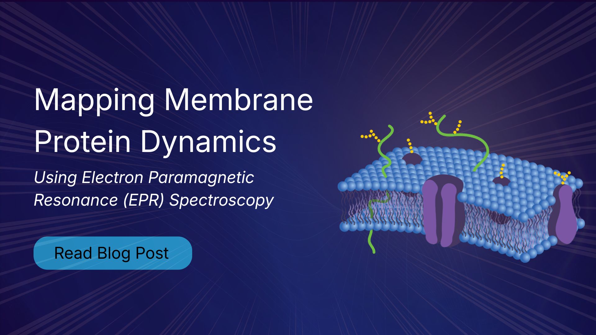
It is well-known that membrane proteins are some of the most important drug targets in biology. These proteins are embedded within or associated with the lipid bilayer that makes up a cell membrane, regulating what passes into and out of the cell while maintaining its internal environment. Membrane proteins mediate signal passage, control the influx of nutrients and the output of waste, and can initiate communication between cells. Because membrane proteins have a role in so many vital physiological processes, that also means that they are often deeply associated with major diseases, including cancers, cardiovascular disease, neurological disorders, and infectious diseases. It also follows that membrane proteins are a popular target for drug discovery programs, and that many drugs we use today act on membrane proteins such as receptors, ion channels, and transporters. However, some of the very properties that make membrane proteins so important in drug discovery can also make them quite difficult to study.
Why Are Membrane Proteins Difficult to Study?
In addition to being a very important class of biomolecules, membrane proteins are also among the most experimentally challenging to investigate. Their inherent dependence on a lipid membrane presents the first fundamental difficulty. Unlike soluble proteins, membrane proteins tend to become unstable when removed from their native lipid environment, and even in rare cases in which a membrane protein may exhibit stability outside a membrane, any measurements made may not be indicative of its behavior in its native environment. To stabilize such proteins and represent the cell membrane, researchers typically employ some sort of membrane mimetic. These are often mixtures of detergents or lipids that form micelles, vesicles, or bilayers. This adds additional complexity to protein expression and purification workflows.
The second fundamental challenge to studying membrane proteins is that they exist as dynamic conformational ensembles and continuously shift between functional states. Many of these states are short-lived or only weakly populated. Ion channels, for example, depend on tightly regulated transitions between open, closed, and inactivated states. Receptor tyrosine kinases (RTKs) rely on dimerization and structural rearrangements to propagate signals that control growth and division. Adhesion molecules (CAMs) coordinate how cells stick, migrate, and organize tissues. Transporters manage chemical gradients that sustain the cellular life cycle. Detailed characterizations of these systems and their mechanisms can prove challenging, as many of key tools in structural biology, such as X-ray crystallography and cryo-electron microscopy, provide structural snapshots in place of the full dynamic picture. NMR spectroscopy can provide some dynamic information but is often limited by protein size and complexity. As a result, researchers are often left with high-resolution protein structures that do not fully capture membrane protein dynamics in functional motion.
How does Electron Paramagnetic Resonance Work?
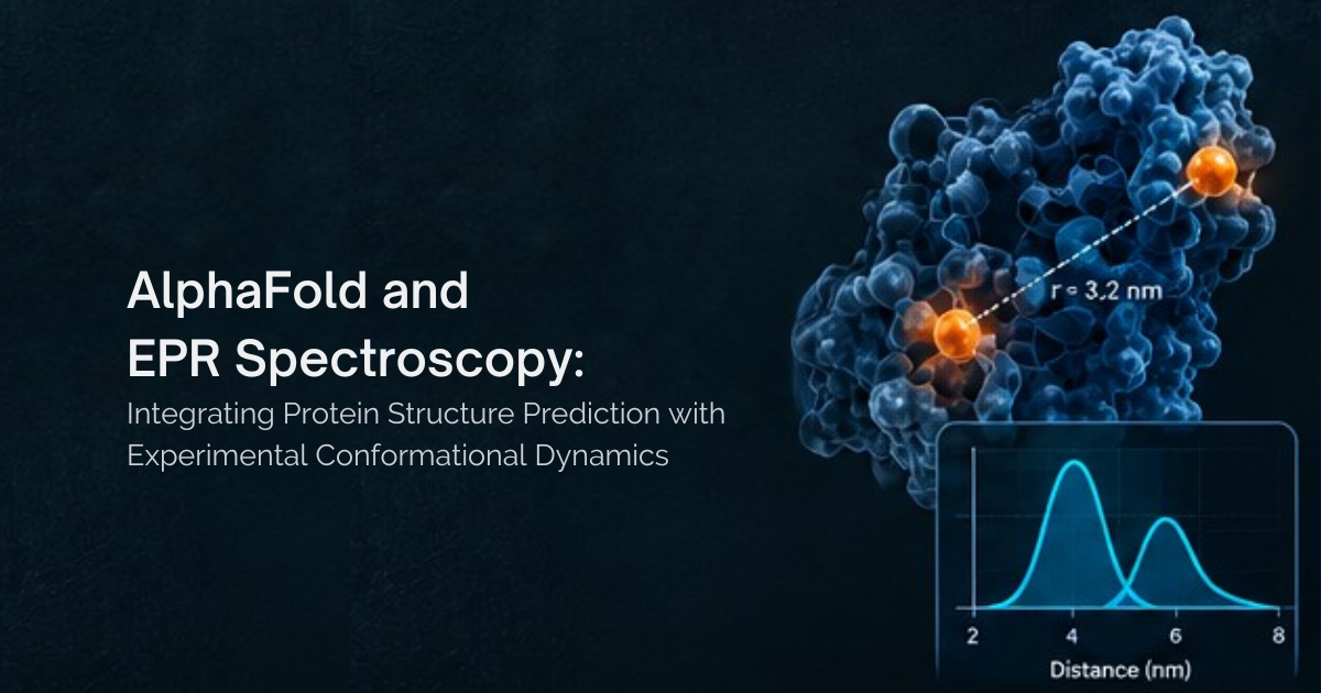
Electron paramagnetic resonance (EPR) spectroscopy is a biophysical technique used to study molecular motion by measuring nanoscale distance distributions between paramagnetic spin labels within a biomolecule. These distance distributions are probability distributions of the distances between spin labeled sites and represent the full conformational ensemble of the system. Multiple concurrent conformations would be represented by multiple peaks in a distance distribution. Changes in conformational state in response to stimuli such as pH change or ligand binding can be tracked through differences in distance distribution. These EPR measurements are easily done on proteins within their membrane mimetic and can even be done on proteins implanted directly within an actual cell membrane.
Applications of EPR in membrane protein research
GPCRs (G protein-coupled receptors) Conformational Dynamics
G protein-coupled receptors (GPCRs) are involved in almost every major physiological process, including signaling, immune system response, and sensory perception, and do so via complex conformational modulation and interactions with ligands and G proteins. Mapping the complex conformational landscape of GPCRs is a challenge for methods such as X-ray diffraction and NMR due to limitations in native membrane conditions and protein size. In response, EPR has become a key method for studying GPCR conformational dynamics that overcomes the above-mentioned limitations of other methods by using minimally perturbing spin labels having no effective limit on macromolecular size.
Case study: G Protein Coupling Specificity in the β2 Adrenergic Receptor Revealed by DEER Spectroscopy
β2 adrenergic receptors are GPCRs expressed in cardiac myocytes and play essential roles in the regulation of cardiac function by the sympathetic nervous system. This receptor primarily couples to the stimulatory G protein Gαs, which increases heart rate and contractility, but it can also activate the inhibitory G protein Gαi, which can counteract Gαs-mediated effects and activate distinct downstream pathways. However, how the receptor selectively couples to different G proteins, and how certain ligands promote one signaling pathway over another, is still fragmentary.
Using DEER spectroscopy, researchers investigated the conformational dynamics of the β2 adrenergic receptor in the presence of different ligands and G protein subtypes. The results showed that the receptor exists in multiple conformations even without ligand, and that differences in receptor conformational states contribute to G protein specificity. The results also support a conformational selection model in which the Gαi-biased ligand LM189 stabilizes a conformation that resembles the Gαi-bound state, facilitating Gi recruitment. This study shows that small changes in receptor dynamics are important for biased signaling, and that DEER spectroscopy can detect these conformational differences in membrane proteins and provide insight into how receptor structure influences G protein selectivity.

Ion Transporters and Membrane Dynamics
Ion transporters are best known for their role in controlling the movement of ions across biological membranes and maintaining electrochemical gradients essential for cellular function. They cycle between outward-facing and inward-facing conformations, which in turn enables substrate transport and coupling to cellular energy and signaling processes. While this conformational flexibility is essential for function, it makes it difficult to capture using traditional structural methods and as a result EPR spectroscopy has emerged as a powerful technique for studying transporter protein dynamics.
Case study: How glutamate transporters move molecules across membranes
The sodium and aspartate symporter GltPh is a membrane transport protein that serves as a structural model for human glutamate transporters, which regulate neurotransmitter levels in brain synapses. These transporters are essential for maintaining proper neuronal signaling and are therefore highly relevant to neurological disease and drug discovery.
Using Double Electron-Electron Resonance (DEER) spectroscopy, performed across multiple transport-domain sites, under different functional conditions (apo, Na⁺/aspartate-bound, and inhibitor-bound) and in two environments (detergent micelles and lipid bilayers), researchers showed that GltPh protomers exist in both outward-facing and inward-facing conformations, in both environments and in both ligand-bound and unbound states.
Need to know how EPR spectroscopy fits into your membrane protein research?
Speak with our application scientist.
Further Reading

It is evident that electron paramagnetic resonance (EPR) spectroscopy has become a key tool for studying membrane proteins in structure-based drug discovery across receptors, transporters, and ion channels, particularly where function depends on dynamic conformational changes. The unique strength of EPR spectroscopy lies in its ability to capture conformational ensembles in native-like membrane environments, including states that are transient, heterogeneous, or not readily resolved by crystallography or cryo-EM, or that are difficult to access due to size. This makes EPR spectroscopy a valuable approach for linking protein structure with dynamics and function in physiologically and pharmacologically relevant systems.