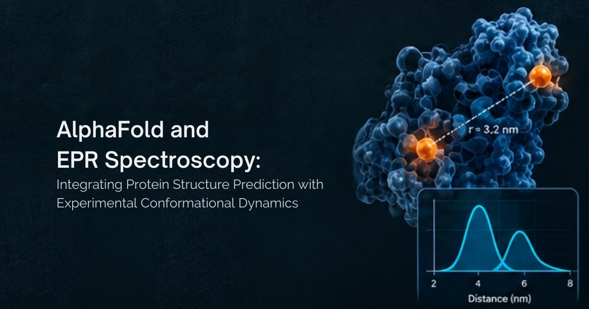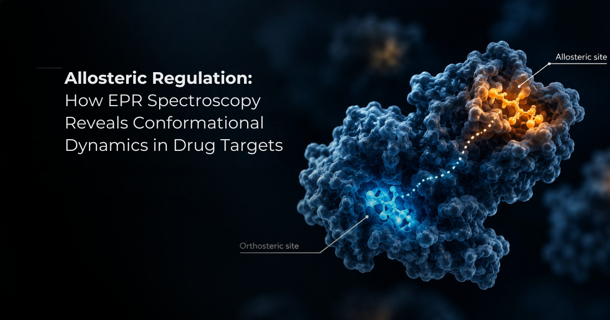
Structure-based drug design uses the three-dimensional structure of a target protein to identify functional binding sites and design drug molecules that complement their shape, size, and electrostatic properties. This approach has enabled major advances in medicine, including HIV protease inhibitors, antibiotics, and targeted cancer therapies such as imatinib. The structural features of a protein’s functional site are obtained through experimental techniques or computational protein modeling and provide the foundation for structure-based drug design. However, while protein structure is highly influential on protein function, it is their dynamic nature that truly dictates physiological functions on a mechanistic level. In this blog post, we will look at the key principles of protein structure and dynamics, why protein conformational dynamics matter in drug discovery and examine how electron paramagnetic resonance spectroscopy contributes to the rational design of effective drug molecules.
The Role of Protein Structures in Drug Design and Discovery
Advances in recombinant DNA technology, genome sequencing, and structural genomics have expanded our understanding of how proteins function and how their activities can be modulated for therapeutic benefit. Proteins are undoubtedly important to nearly every cellular process, from nucleic acid synthesis and energy production to lipid metabolism, and their function is closely linked to their structure, dynamics, and interactions with other biomolecules. At the molecular level, proteins are described across four hierarchical levels: primary, secondary, tertiary, and quaternary, with their conformations influenced by their environment and interactions within the crowded cellular milieu. As a result, changes in protein structure, dynamics, or assembly can disrupt molecular recognition and contribute to disease. These properties also make proteins highly valuable targets for drug discovery, with receptors, enzymes, ion channels, and transporters accounting for many established drug targets.
Notably, three-dimensional biomolecular structures provide insight into functional sites and drug-binding pockets. This structural information forms an important foundation for structure-based drug design. Our basic biochemistry knowledge reminds us that the amino acid sequence determines how a protein folds through hydrogen bonding and other non-covalent interactions, forming secondary structures such as α-helices and β-sheets that assemble into a stable three-dimensional fold. Once formed, this fold organizes the protein residues according to their physicochemical properties. Consequently, in globular proteins, hydrophobic residues are generally buried within the core, while polar residues are exposed to the surface. This arrangement, in turn, positions functional residues precisely within active or binding sites, creating the physicochemical environment required for biological activity.
Experimentally, protein structures can be determined using techniques such as X-ray crystallography, nuclear magnetic resonance (NMR), and cryo-electron microscopy (cryo-EM), or predicted computationally. While these approaches have generated an extensive collection of structures in the Protein Data Bank (PDB), the number of proteins with known sequences continues to far exceed those with experimentally determined structures. Intrinsically dynamic or very large proteins can also present challenges for conventional structural methods. To bridge this gap, approaches, including electron paramagnetic resonance (EPR) spectroscopy and computational modeling, are employed to provide a more complete view of protein structure and dynamics.
Learn more about electron paramagnetic resonance (EPR) spectroscopy here: What is EPR Spectroscopy? Principles, Spin Labeling, and DEER in Proteins - High Q Technologies Inc. | HighQ Technologies
How Structure-Based Drug Design Uses Protein Structures
Protein function depends largely on interactions with other molecules, or ligands, including proteins, DNA, RNA, ions, neurotransmitters, and other small molecules. These interactions, in turn, are governed by the structural and electrostatic complementarity between the interacting surfaces. In proteins, these features are determined by the three-dimensional arrangement of amino acid residues. Structure-based drug design (SBDD) then uses this information to develop molecules that bind selectively to a protein target and modulate its function. For drugs targeting orthosteric sites, the goal is often to reproduce key structural and electrostatic features of the endogenous ligand while optimizing interactions with the binding pocket. In this context, detailed knowledge of the pocket enables researchers to select and position chemical groups that maximize binding affinity, selectivity, and functional activity.
Examples of Structure-Based Drug Design
Drug discovery is a lengthy and resource-intensive process, typically progressing from target identification and validation through lead discovery, preclinical development, clinical trials, and regulatory approval. Thus, SBDD has become an important component of this process by using structural and computational information to accelerate the identification and optimization of potential drug candidates.
Once a target has been validated, researchers identify molecules capable of binding to and modulating the target. To support this, molecular docking is commonly used to screen large compound libraries and prioritize molecules with favorable interactions with the target binding site. Reliable three-dimensional protein structures obtained through techniques such as X-ray crystallography, NMR, and cryo-electron microscopy provide an important foundation for these studies.
Several landmark medicines show the impact of SBDD. HIV-1 protease inhibitors, including saquinavir and indinavir, represent some of the earliest successful applications of structure-guided drug discovery. Another major milestone was the development of imatinib, a BCR-ABL kinase inhibitor approved in 2001 for the treatment of chronic myeloid leukemia. Its discovery demonstrated how structural information about a protein’s conformational states can reveal opportunities to achieve greater target selectivity. In particular, targeting the less-conserved inactive conformation of the kinase helped guide the development of a highly selective therapeutic.
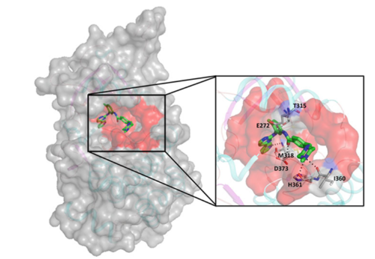
Fig 1: Imatinib bound to the inactive conformation of ABL tyrosine kinase. ABL is shown as a transparent grey surface and imatinib as green sticks, with the binding site highlighted in red. The enlarged view shows the key protein residues involved in interactions with imatinib.
Image source: Arangasamy, Yazhini & Chakraborti, Sohini & Srinivasan, Narayanaswamy. (2021). Protein Structure, Dynamics and Assembly: Implications for Drug Discovery. 10.1007/978-981-15-8936-2_5.
Why Does Protein Dynamics Matter in Drug Discovery?
Protein’s biological function depends not only on a protein structure, but also on the inherently dynamic nature of proteins to adopt multiple conformations. Moreover, increasing evidence shows that these conformational dynamics play an important role in molecular recognition and drug binding. Classical models of molecular recognition, including the “lock-and-key” and “induced-fit” models, have evolved to incorporate conformational selection, in which a ligand preferentially binds to a pre-existing conformation of a dynamic protein. This principle applies broadly to protein–small molecule, protein–protein, and protein–nucleic acid interactions. You might wonder, why do you need to understand these conformational states of proteins? Allosteric sites, for example, can be targeted to alter protein activity indirectly by stabilizing specific conformations, but allosteric sites are not always evident. A well-known example is Imatinib, mentioned earlier. This drug preferentially binds the inactive conformation of ABL kinase. This clearly affirms that the knowledge of conformational dynamics of a target protein indeed aids target selectivity.
Another important example of the role of dynamics in drug action is membrane proteins. N-methyl-D-aspartate (NMDA) receptors are glutamate-gated ion channels that switch between closed and open states to regulate calcium ion flow in neurons. Given how central NMDA receptors are to healthy brain function, it's not surprising that schizophrenia, Alzheimer's, Huntington's, Parkinson's, and ALS have all been linked to NMDA receptor dysfunction. Under pathological conditions, prolonged receptor activation can contribute to neuronal damage. Memantine (uncompetitive, voltage-dependent NMDA receptor antagonist) preferentially targets the open state of the NMDA receptor, reducing excessive activity while preserving normal signaling. This led it to it becoming a successful therapeutic intervention.
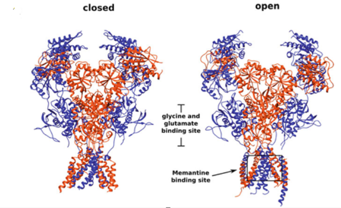
Fig 2: NMDA receptor in its ‘closed’ (PDB: 4TLL; Lee et al. 2014) and ‘open’ (PDB: 6IRA; Zhang et al. 2018) conformations. GluN1 and GluN2 subunits are shown in blue and orange, respectively, with the memantine binding site highlighted by the box.
Image source: Arangasamy, Yazhini & Chakraborti, Sohini & Srinivasan, Narayanaswamy. (2021). Protein Structure, Dynamics and Assembly: Implications for Drug Discovery. 10.1007/978-981-15-8936-2_5.
In a similar vein, G protein-coupled receptors (GPCRs) exist in multiple conformational states that regulate signal transmission. Movements of their transmembrane helices and extracellular and intracellular regions can influence ligand binding and create alternative sites for allosteric modulators. These dynamic properties are important in the design of selective GPCR-targeted therapies.
Techniques for Studying Protein Structure and Dynamics
Electron Paramagnetic Resonance (EPR) spectroscopy measures the behavior of unpaired electrons introduced at specific sites in a protein, typically through site-directed spin labeling. It basically involves attaching a paramagnetic spin label to selected positions, while monitoring the changes in the local environment, mobility, and distances between labeled sites on the nanometer scale. As a result, distance distributions and populations of different conformational states are revealed, offering an avenue to gain insights into the dynamic structural ensembles that underlie protein function. It’s no wonder that EPR spectroscopy has been applied to proteins that are difficult to study using conventional structural approaches, including flexible proteins, intrinsically disordered regions, membrane proteins, transient complexes, and aggregation-prone systems.
EPR distance constraints are excellent complements to the other techniques used to study protein structure. X-ray crystallography structures represent an average of protein conformations. Also, ensemble structures from solution NMR technique provide information on large scale dynamics of proteins, but NMR is limited by size. Other approaches, such as smFRET provide valuable information on protein dynamics at the single-molecule level. Advances in cryo-EM have also made it possible to study large-scale conformational changes in proteins and molecular assemblies in greater detail. Combining these methods with Pulsed EPR methods such as double electron-electron resonance (DEER) has made it significantly easier to probe protein dynamics and conformational ensembles.
How EPR Spectroscopy Reveals Membrane Protein Structure and Dynamics | HighQ Technologies

The outcome of protein–protein and protein–ligand interactions depend on the interplay between sequence, structure, and dynamics. As mentioned earlier, through the transitioning between different conformations, proteins can respond to their environment and regulate interactions with binding partners, making dynamics an important factor in drug discovery. These conformational changes can reveal cryptic binding sites and create new opportunities for targeting difficult-to-drug proteins. In addition, protein dynamics play a key role in allosteric signaling. As understanding the total conformational ensemble of such vital processes and systems continues to gain importance, EPR (Pulsed EPR Spectroscopy) presents a powerful and accessible strategy to characterize the conformational dynamics of biological systems.