Previously I provided a high-level view of the fundamentals of EPR-based distance measurements. Today, and in future iterations, I will dig deeper into those distance methods, focusing on specific techniques, their strengths, drawbacks, and considerations. Each instance produces similar results – a distance distribution between two unpaired electron spins – but differ in implementation and optimization. Understanding the various factors at play within these methods is crucial to pair the optimal experiment with a given sample and environment.
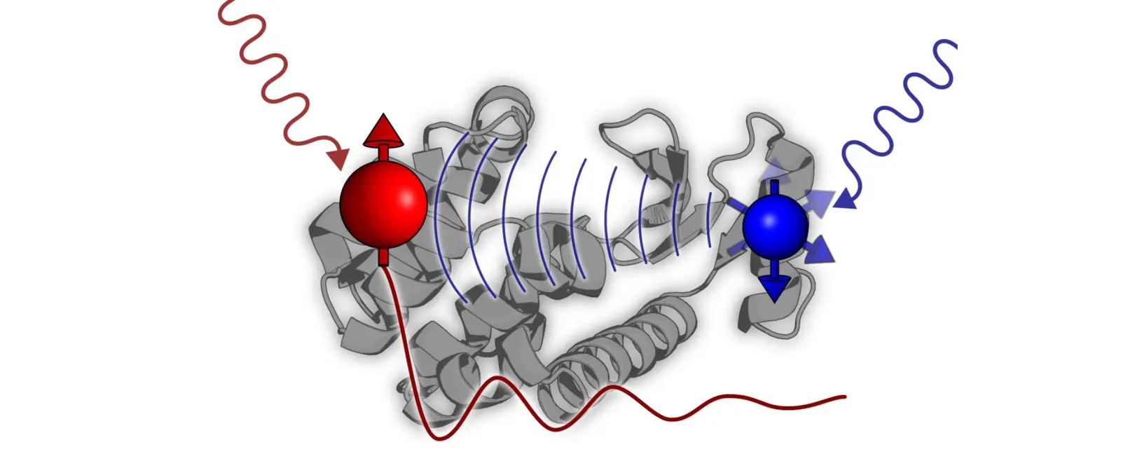
This first installment will focus on the most ubiquitous distance method in EPR – Double Electron -Electron Resonance, or DEER, and sometimes PELDOR (Pulsed Electron Double Resonance). This experiment utilizes two microwave frequencies in a pump/probe scheme: One microwave frequency is used to probe, or monitor, a set of spins within the system, denoted ‘A’ spins. The second ‘pump’ frequency is used to flip a separate set of spins, ‘B’ spins. Because each individual spin has an intrinsic magnetic moment, the flipping of the ‘B’ spins slightly alters the overall magnetic field experienced by the ‘A’ spins, which impacts the amplitude of the measured signal. By varying the time at which the ‘B’ spins are flipped, we observe a modulation of the monitored ‘A’ spins at a specific frequency, which is related to the inverse of the inter-spin distance, cubed (1/r3).
The DEER experiment has several specific iterations, distinguished by the number of pulses used in the sequence. Without getting too deep into the weeds, the initial incarnation of DEER was introduced by Milov et. al. and featured three pulses – two probe and one pump. While initially successful, this sequence suffered from dead time, wherein the entire dipolar trace could not be collected, which led to ambiguity in the overall analysis. This issue was remedied in the most widely used version of DEER, and the most widely used EPR distance technique in general, the 4-pulse DEER, introduced by Pannier et. al. This version of the experiment removes dead time from the sequence entirely, allowing robust and unambiguous data analysis. Further variations of DEER have been developed for more niche applications, such as 5- and 7- pulse DEER, which make use of a Carr-Purcell pulse sequence to mitigate relaxation effects on the observed signal and thus extend the range of measurable distances. While successful in their goal, these techniques are more complicated to set up and require heavier post-processing to extract useable distance data. LiDEER has also been developed, replacing one spin label with a photoactivable porphyrin group, and using laser excitation in place of a second microwave frequency to pump the system. This technique requires orthogonal labels, and a spectrometer with an optical window for laser excitation.
In EPR, DEER is currently as close to a one-size-fits-all distance technique as exists, although there are systems where other techniques would be better suited, and a few in which DEER is simply not feasible. That said, DEER is widely applicable, reliable, and relatively straightforward to analyze. From a hardware standpoint, DEER requires a system capable of producing two different microwave frequencies, and a resonator with the bandwidth to accommodate them. DEER works very well on nitroxide spin labels, as they are spectrally broad enough that the excitation profiles of the two frequencies can be separated but narrow enough that most of the spectrum can still be excited. DEER is also easily applied to metal ions, although with most metals being significantly spectrally broader than nitroxides, sensitivity will suffer. However, DEER is poorly suited to measure Trityl spin labels, as their spectra is so narrow that the two microwave frequencies typically cannot be separated, although exceptions exist.
Regarding data collection, most EPR distance techniques require two experimental addendums – phase cycling, which eliminates unwanted signals from the final data, and nuclear modulation averaging, which subtracts the effects of neighboring nuclei on the measured electrons. DEER requires only modest application of these processes to yield quality data, whereas other techniques require more complex schemes. Analysis of DEER is relatively robust as well. The measured signal in EPR distance measurements typically displays a background signal that must be accounted for either before or during data analysis. For DEER, this background is well known as a standard exponential decay in most cases, easing the data analysis workflow (for other techniques, this is not always the case and is a major area of interest in the development of new sequences). Further analysis of this background decay can also be used to estimate local sample concentration and spin labeling efficiency.

DEER is an incredibly useful technique in EPR – and biophysics in general – that has seen great success and wide adoption in the field. A reliable and well-tested method, DEER data has resolved many important biological distances and paved the way for further studies. Without DEER in its various incarnations, the field of EPR distance measurements would likely look starkly different today. Follow along in the coming months as I continue to explore EPR distance techniques. ‘Til next time!