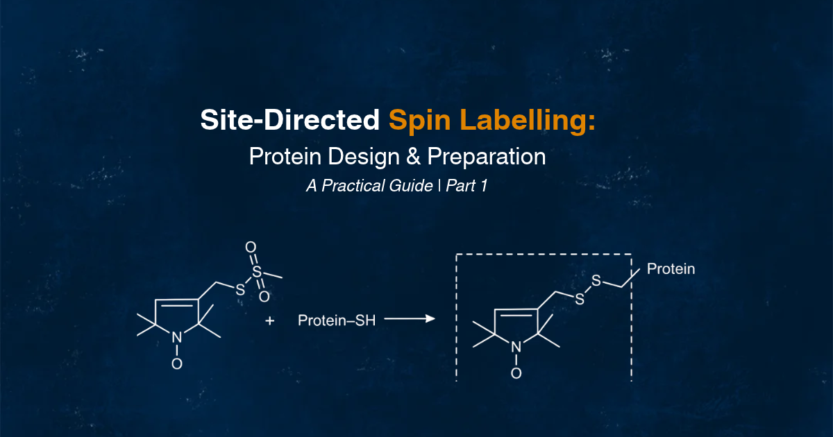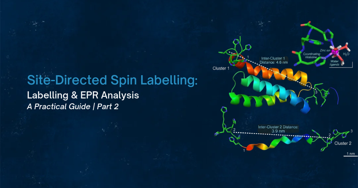
In Part 1 of this guide, we explored the fundamentals of site-directed spin labelling (SDSL), including how to select suitable spin-label positions and prepare proteins for labelling.
In Part 2, we continue with the practical workflow, covering the spin-labelling process, how to verify labelling efficiency, and how to troubleshoot common challenges.
If you haven't read Part 1 yet, you can find it here: [Site-Directed Spin Labelling (SDSL): A Practical Guide to Protein Design and Preparation].
How do you prepare and perform the spin-labelling reaction?
Before adding MTSL, the engineered cysteine must be available in its reduced thiol form. If the cysteine has oxidized during protein overexpression via a post-translational modification, reducing agents such as dithiothreitol (DTT) or TCEP may be used during sample preparation to remove this modification. However, reducing agents must be removed before adding MTSL because they heavily interfere with binding of the spin-label reagent. A size-exclusion or desalting column, dialysis or another suitable method can be used to remove the reducing agent and transfer the protein into a compatible labelling buffer.
A typical MTSL labelling workflow involves:
- Prepare the protein: Transfer the purified cysteine-containing protein into a suitable labelling buffer.
- Make the cysteine thiol available: Reduce the cysteine if necessary, using an appropriate reducing agent.
- Remove the reducing agent: Exchange the buffer or desalt the protein before adding MTSL.
- Prepare the spin label: Prepare an MTSL stock solution (typically in organic solvent) according to the reagent manufacturer's instructions and the established experimental protocol.
- Perform the reaction: Add an appropriate molar excess of MTSL relative to the available cysteine sites (typically 5 – 20 fold molar excess over cysteines) Never exceed 1 – 2 % v/v of organic solvent in your labelling reaction solution as this may hurt your protein integrity.
- Incubate: Allow the reaction to proceed under conditions suitable for the particular protein.
- Remove unreacted label: Use desalting, size-exclusion chromatography, dialysis or another suitable purification method.
Several factors influence the labelling reaction. Buffer composition and pH affect cysteine reactivity and the stability of both the protein and spin label. Reducing-agent removal is important because reducing agents typically reduce labelling efficiency. Protein concentration can influence aggregation and reaction conditions as well. Finally, temperature and incubation time affect reaction kinetics and protein stability, so both should be balanced out and adjusted to achieve efficient labelling without compromising the protein integrity. There is no single universal MTSL labelling recipe that fits all proteins. Reaction conditions should be optimized for each protein rather than transferred directly from one to another system – the general procedure remains similar though. The resulting MTSL–cysteine linkage is reduction-sensitive. If the experiment requires strongly reducing conditions, a different labelling chemistry may be more appropriate.
How do you verify labelling efficiency and protein quality?
A completed labelling reaction does not automatically mean that the sample is ready for EPR measurements. You need to establish three things: whether the label has attached, how much label is present and whether the protein remains suitable for the intended experiment.
1. Confirm that the spin label has attached
Mass spectrometry can help confirm the expected molecular mass change following spin-label attachment. It can also reveal incomplete or heterogeneous labelling. For MTSL, the expected mass increment is approximately 184 Da per modified cysteine. Accordingly, the exact value should be calculated for each spin label in consideration so that it can be identified via mass spectrometry.
2. Determine spin concentration and labelling efficiency
Spin counting in CW-EPR can be used to estimate the number of paramagnetic labels relative to the protein concentration. Comparing the measured spin concentration with the expected number of labels per protein molecule is the key for assessing labelling efficiency. For a doubly labelled construct, for example, the expected number of spins per fully labelled protein is two. Incomplete labelling reduces the modulation depth in PDS and the therefore the quality of the resulting measurements.
3. Check protein integrity and biological activity
The labelled protein should be assessed to confirm that it retains its expected structural and functional properties. Depending on the protein, this may include comparing its secondary or tertiary structure with the unlabelled protein, assessing stability and aggregation, measuring ligand binding, and performing an activity assay or another relevant functional test. The wild-type protein and unlabelled mutant are useful controls for distinguishing effects caused by the cysteine mutation from those caused by the attached spin label. This is typically done via ultraviolet circular dichroism (UV-CD) spectroscopy.
4. Check for residual free spin label
Unreacted spin label must be removed before pulsed dipolar EPR measurements. Free label can contribute to the background signal and compromise data quality. CW-EPR spectra can help identify residual unbound label, while appropriate purification methods or repetitive/iterative steps should be used to remove it. The EPR community guidelines specifically emphasize the importance of knowing protein concentration and labelling efficiency, removing free label and verifying that protein structure and function have not been compromised.
What can go wrong during SDSL, and how can you troubleshoot it?
Several experimental issues can affect labelling efficiency and EPR data quality. Understanding their possible causes helps you identify problems before interpreting the results.
Low labelling efficiency
Possible causes include inaccessible or oxidized cysteine residues, residual reducing agents and incompatible reaction conditions. Check cysteine availability (e.g. via Ellman’s test), buffer composition and reaction conditions. Consider whether the selected labelling site is sufficiently accessible.
Unwanted or non-specific labelling
Native cysteine residues or other unintended reactive sites can produce surplus labelling sites. Review the protein sequence and construct design. Compare the results with appropriate controls, such as wild-type protein or a cysteine-free construct where suitable and feasible.
Protein precipitation or aggregation
Protein instability can be caused by various effects. Self-aggregation via molecular crowding or poor buffer conditions may cause precipitation during sample preparation or labelling. Carefully assess conditions that enhance protein stability before and after the labelling procedure. Consider whether buffer composition, temperature or the selected membrane-mimetic environment needs optimization.
Weak dipolar modulation
Incomplete labelling, insufficient spin concentration or inappropriate sample conditions can reduce the observed modulation depth of the dipolar signal. Check protein concentration, labelling efficiency and sample preparation. Also consider whether the expected spin–spin distance falls within the measurement range of the experimental setup. If you have to work at low concentrations, low labelling efficiencies and/or long distances, always use perdeuterated buffers and cryoprotectants.
Broad or multimodal distance distributions
These can arise from genuine conformational heterogeneity, spin-label rotamers or other computational artifacts. Consider structural models, label flexibility, sample quality and data-processing limitations before attributing the distribution entirely to protein dynamics. It is advisable to consult multiple software packages in a comparative fashion if results vary or remain inconsistent. The EPR community guidelines suggest careful and transparent reporting of experimental and analytical procedures that are used.
You can also use our Site-Directed Spin Labelling Checklist as a practical reference throughout the process. Download checklist.
Frequently asked questions about SDSL
Should spin label sites be solvent exposed?
For all distance and mobility experiments on globular or membrane proteins: yes, a solvent-exposed labelling site is the safest choice. Additional care has to be taken in case a target protein forms oligomers or assemblies with other proteins and putative surface regions may become buried upon association. Make sure you have sufficient structural information available for making a good educated guess where areas might become covered.
Can you spin label a protein without cysteine?
Yes, you can spin label a protein without cysteine by using other residues or engineered motifs as the attachment point. Noncanonical amino acids added by genetic code expansion can carry reactive groups for bioorthogonal labeling, tyrosine-reactive labels target native tyrosines, and e.g. Cu(II)-based spin labels bind particularly well to engineered double histidine (dHis) motifs. These different potential labeling routes help to make a decision when a protein has many essential cysteines, or when the stabilized radical on the label has to withstand reactive oxygen species when EPR is performed in-cell.
Does MTSL change protein structure?
MTSL usually has little-to-no effect on protein structure when it is attached at a solvent-exposed site, which is the main reason exposed sites are preferred for SDSL. Labels placed in the protein core, at tertiary contacts or in binding sites can destabilize the fold or change function. Check every labeled mutant with a fold measurement such as UV-CD and with an activity or binding assay before interpreting its EPR spectrum.
Do you need to deuterate samples for PDS?
Deuteration is not always required for PDS, but it lengthens the usable trace and so extends the distance range and significantly improves data quality. Some samples are also intrinsically not accessible to perdeuteration. Deuterating the buffer and cryoprotectant through a buffer exchange is the cheapest step, and High Q's applications team reported in 2026 that it typically reaches above 94% deuteration. Full protein deuteration (>99%) costs much more and is used for obtaining the longest distances, up to about 16 nm.
How close to a binding site can a spin label go?
Keep labels off residues whose side chains touch the ligand, nucleic acid or partner proteins, and place them on the elements you expect to sufficiently move. As a working rule, skip contact residues and their immediate neighbors in sequence, then confirm with a binding or activity assay on the labeled protein. A label one helix turn away from a pocket on the same helix usually reports the movement just as well as a label in the pocket, without the risk of changing ligand binding affinity.
How do you choose spin label pairs for DEER?
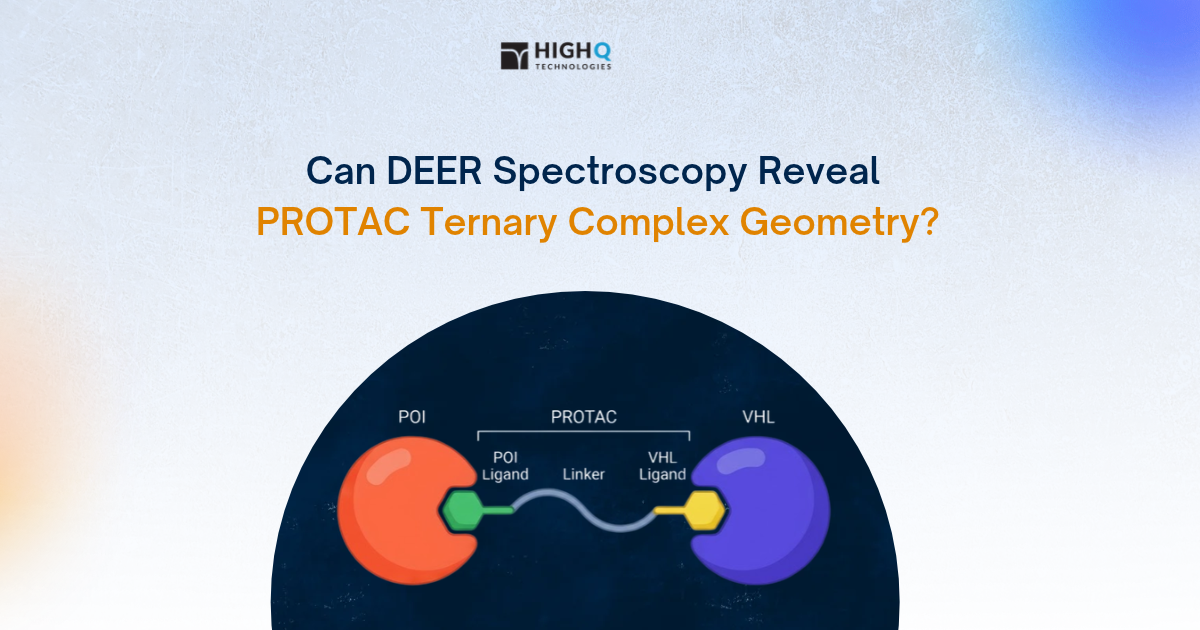
Choose DEER pairs so that the expected label-to-label distance falls inside the range your measurement can resolve, and so the distance clearly changes between the states you want to compare. Put the two labels on different structural elements, such as two different domains or a helix and a loop of the protein, because a pair on one and the same rigid unit will typically not provide a huge distance change when that unit moves.
What distance range can DEER measure?
DEER typically measures distances between about 1.5 and 8 nm, and the upper limit depends on how long the dipolar evolution trace can run before the spin echo has faded away. On High Q Technologies' FATHOM® EPR spectrometer, the reliable range for protonated aqueous samples is typically 1.8 to 6.0 nm with a 3 to 4 µs trace.
How do you predict spin label positions before mutagenesis?
Rotamer library modeling and screening predicts where a label will sit at each candidate site and what distance distribution a pair will give, before you spend your time on cloning and expression. Also, the primary, secondary and tertiary structure gives us a clue which type of labeling approach is best suited.
How many spin label pairs do you need?
One well-chosen pair can confirm a single hinge motion, but locating a domain or building a model usually takes several pairs that share anchor sites. The amount of chosen spin pairs solely depends on the required spatial resolution and topology of the dynamic effect under consideration.
Which spin labels work for in-cell EPR?
In-cell EPR needs labels with linkages and radicals that withstand a highly reducing environment, so site choice and label choice have to be planned together. The cytosol cleaves disulfide bonds and reduces nitroxides to EPR-silent species, which is why in-cell studies use cleavage-resistant linkers,and stabilized radicals such as Gd(III) complexes or trityl radicals.
How do you spin label DNA and RNA?
In DNA and RNA, spin labels are placed via reactive handles at desired oligonucleotide sites that are inserted during synthesis, so you rather choose a specific nucleotide position and an appropriate click-chemistry approach instead of an amino acid residue to replace.
References
- de Vera IMS, Blackburn ME, Galiano L, Fanucci GE. Pulsed EPR distance measurements in soluble proteins by site-directed spin labeling (SDSL). Curr Protoc Protein Sci. 2013;74:17.17.1–17.17.29. doi: 10.1002/0471140864.ps1717s74.
- Altenbach C, López CJ, Hideg K, Hubbell WL. Exploring structure, dynamics, and topology of nitroxide spin-labeled proteins using continuous-wave electron paramagnetic resonance spectroscopy. Methods Enzymol. 2015;564:59–100. doi: 10.1016/bs.mie.2015.08.006.
- Schiemann O, Heubach CA, Abdullin D, Ackermann K, Azarkh M, Bagryanskaya EG, et al. Benchmark test and guidelines for DEER/PELDOR experiments on nitroxide-labeled biomolecules. J Am Chem Soc. 2021;143(43):17875–17890. doi: 10.1021/jacs.1c07371.
- Tessmer MH, Stoll S. Protein modeling with DEER spectroscopy. Annu Rev Biophys. 2025;54:35–57. doi: 10.1146/annurev-biophys-030524-013431.
- Sahu ID, Lorigan GA. Site-directed spin labeling EPR for studying membrane proteins. Biomed Res Int. 2018;2018:3248289. doi: 10.1155/2018/3248289.
- Drescher M. EPR in protein science: intrinsically disordered proteins. Top Curr Chem. 2012;321:91–119. doi: 10.1007/128_2011_235.
- Hubbell WL, Mchaourab HS, Altenbach C, Lietzow MA. Watching proteins move using site-directed spin labeling. Structure. 1996;4(7):779–783. doi: 10.1016/S0969-2126(96)00085-8.
- Jeschke, G. (2018). MMM: A toolbox for integrative structure modeling. Protein Science, 27(1), 76-85.