What is site-directed spin labelling, and why is it used in protein research?
Site-directed spin labelling (SDSL) is a technique that combines protein engineering with electron paramagnetic resonance (EPR) spectroscopy to investigate protein structure, conformational changes and dynamics. The approach involves introducing a paramagnetic spin label at a selected position in a protein. The label contains an unpaired electron that can be detected by EPR, allowing researchers to investigate its local environment or measure distances between two labelled sites.
Most proteins do not contain naturally occurring paramagnetic centres suitable for these measurements. SDSL addresses this limitation by introducing a paramagnetic probe at a specific location. The most established approach uses site-directed mutagenesis to introduce a cysteine residue, commonly followed by attachment of a thiol-reactive nitroxide spin label. Depending on the experimental design, SDSL-EPR can provide information about:
- Local protein structure and side-chain mobility.
- Protein backbone flexibility and solvent accessibility.
- Relative movements between protein domains.
- Protein–protein interactions and complex formation.
- Structural ensembles and conformational heterogeneity.
- Conformational changes between different functional states (pH, light, ligand, etc.).
SDSL is particularly useful when researchers need information about protein behaviour in solution, including structural changes that may not be apparent from a single static structure. It complements methods such as X-ray crystallography, cryo-EM and NMR by providing site-specific information about protein dynamics and distance constraints.
How does a spin label attach to a protein?
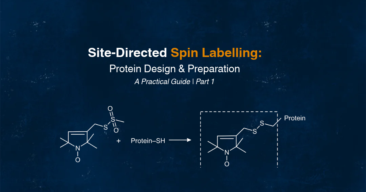
In conventional cysteine-based SDSL, an engineered cysteine residue provides the attachment point for a thiol-reactive spin label. MTSL (methanethiosulfonate spin label) is one of the most widely used nitroxide spin labels. It reacts with the cysteine thiol group through thiol–disulfide exchange, forming a disulfide-linked paramagnetic side chain commonly referred to as R1. As the covalently attached nitroxide contains an unpaired electron, it produces an EPR signal. This chemistry allows researchers to introduce a paramagnetic probe at a selected position without changing the entire protein or relying on a naturally occurring paramagnetic centre.
However, the MTSL–cysteine linkage is a disulfide bond and is sensitive to reducing conditions. This is an important consideration when choosing buffers, preparing samples and designing experiments. Other spin-label chemistries are available:
- Maleimide-based labels: React with cysteine thiols to form thioether linkages, which are generally more resistant to reduction than the MTSL disulfide linkage.
- Iodoacetamide-based labels: Provide another route for modifying cysteine residues for higher pH stability.
- Unnatural amino acid labelling: Introduces alternative reactive groups at selected positions when conventional cysteine labelling is unfavorable.
- Alternative paramagnetic labels: Metal-based spin labels, including gadolinium- or copper-containing species, can be useful in particular for pulsed EPR applications.
The choice of chemistry depends on the protein, the experimental environment and the type of EPR measurement required. Choosing spin-label sites starts with the structural question you want to answer. Are you investigating local protein mobility, the movement of two domains, a protein interaction or a conformational change following ligand binding? The answer will guide where the spin labels should be introduced. For CW-EPR, a single spin label can provide information about local mobility and its surrounding environment. In pulsed dipolar spectroscopy (PDS), two spin labels are introduced at selected positions to measure interspin distances and investigate structural changes, such as the movement of two domains relative to another. When selecting candidate sites, consider the following factors:
- Solvent accessibility: Choose cysteine positions that are accessible to the labelling reagent. Buried residues are usually difficult to label efficiently.
- Structural location: Consider the structural elements involved in the movement or conformational change you want to investigate. Stable regions can provide useful reference points, while flexible regions may be relevant when studying protein dynamics.
- Protein function: Avoid catalytic residues, essential binding interfaces (e.g. for dimerization) and residues involved in important disulfide bonds, unless modifying them is part of the experimental design.
- Structural sensitivity: For PDS, select two positions where the expected structural change is likely to produce a measurable difference in interspin distance.
- Native cysteines: Identify existing cysteine residues that could react with the labelling reagent or contribute to structural disulfide bonds.
- Spin-label flexibility: Consider how the attached spin-label side chains and their rotamers could influence the measured distance distribution. A decisive factor is the distance of the unpaired electron to the C-alpha atoms of the protein backbone.
Crystal structures, cryo-EM structures and computational models can help in identifying candidate positions. Structural visualization tools can be used to examine solvent accessibility, estimate distances between proposed labelling sites and assess whether a label pair is likely to report on the structural change of interest. Where possible, spin-label modelling tools such as MMM or chiLife can help to evaluate most probable label conformations and predicted interspin distance distributions. For example, if ligand binding is expected to change the relative arrangement of two protein domains, one cysteine position could be selected on each domain, preferably in accessible topological regions that are not essential for protein function. After introducing the spin labels, PDS measurements can compare the interspin distance distributions in the absence and presence of the ligand. Once candidate sites have been selected, prepare the cysteine mutants and assess their expression, stability, structural integrity, function and labelling efficiency. These checks help establish whether the labelled protein mutant candidate remains suitable for the planned EPR experiment.
How do you design and prepare a protein for SDSL?
Once candidate positions have been identified, the next step is to prepare the protein construct. In conventional cysteine-based SDSL, site-directed mutagenesis is used to introduce cysteines at the desired positions. Depending on the protein, native cysteine residues may also need to be considered. Some can react with the spin label and produce unwanted labelling, while others form structural disulfide bonds or contribute to protein function. Researchers may therefore replace selected native, non-essential cysteine residues with other amino acids to reduce non-specific labelling. However, this strategy must be evaluated carefully because removing native cysteines can affect protein structure and function. A typical protein preparation workflow involves:
- Designing the cysteine-containing protein construct.
- Introducing the required mutations and verifying the DNA sequence.
- Expressing the recombinant protein in a suitable expression system.
- Purifying the protein using an appropriate method.
- Assessing protein purity, stability and aggregation behaviour.
- Confirming that the engineered protein retains relevant biological activity.
The unlabelled mutant should be characterized before proceeding to spin labelling. This provides a baseline for determining whether subsequent changes are caused by the label rather than by the mutation itself. Membrane proteins require additional attention because their stability and functionality depend on the surrounding membrane environment. One may need to maintain the protein in detergent micelles, lipid nanodiscs or another suitable membrane-mimetic system of appropriate lipid composition. There is no universally suitable membrane-mimetic environment for every protein. The selected system must maintain protein stability and function while remaining compatible with the intended EPR experiment.
Learn more about which spin labels to use here
Planning an SDSL experiment? Use our Site-Directed Spin Labelling Checklist to help guide your protein design and preparation before moving on to the labelling process.
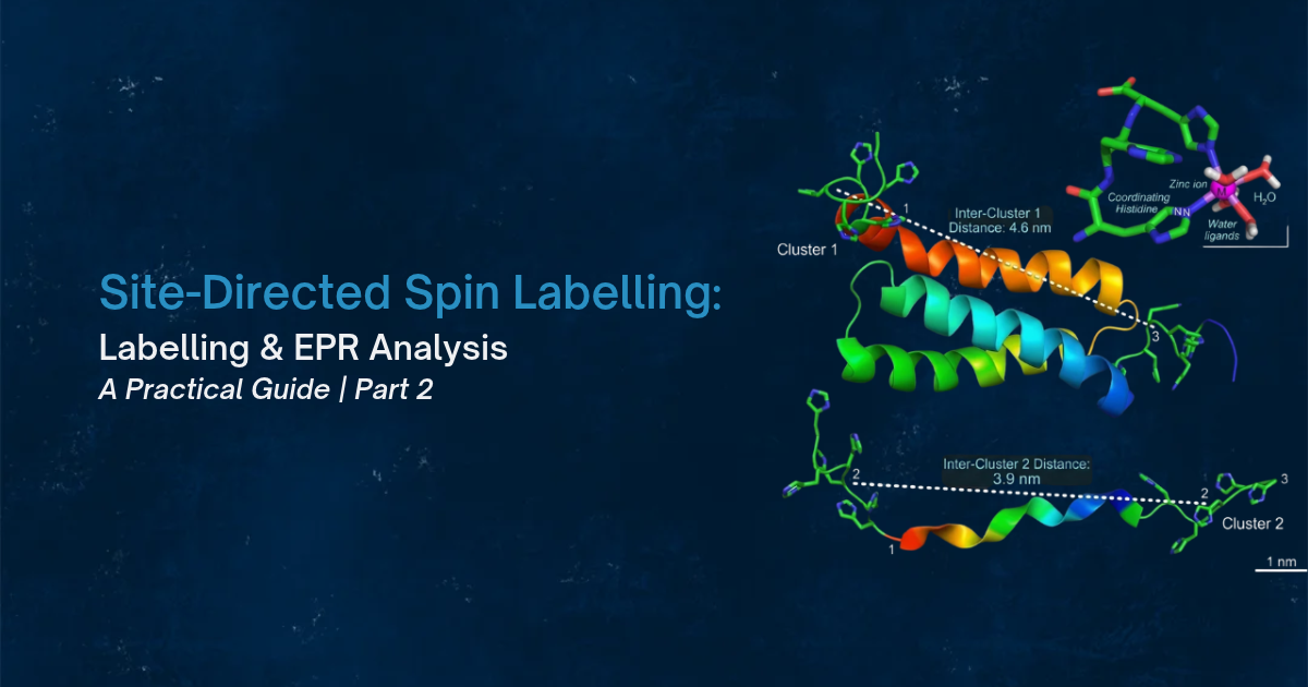
Continue reading: Part 2 – Site-Directed Spin Labelling, EPR Analysis and Troubleshooting.