EPR Spectroscopy for GPCR & Membrane Protein Dynamics
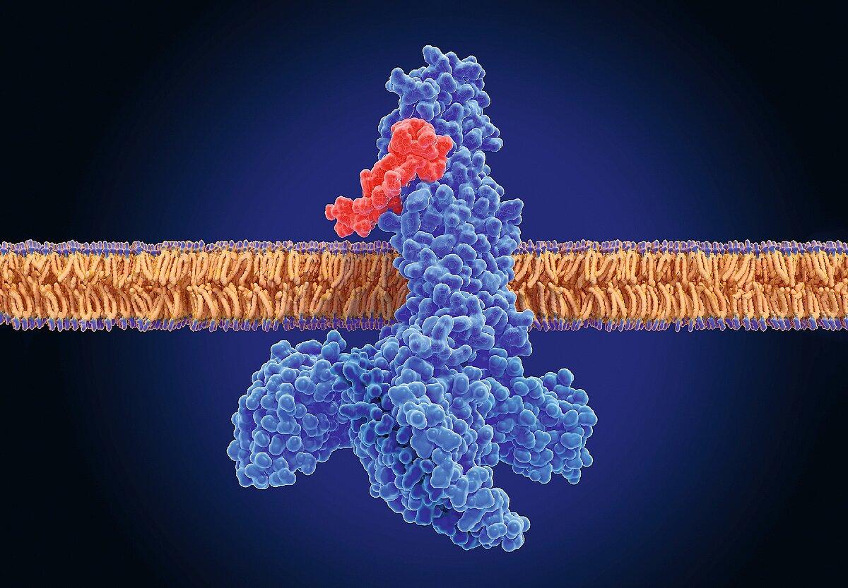
G protein-coupled receptors (GPCRs) are one of the largest families of cell-surface receptors, playing a critical role in cellular signalling and regulating a wide range of biological processes. As dynamic membrane proteins, GPCRs undergo structural and conformational changes in response to ligand binding, influencing how they interact with other proteins and transmit signals.
Electron paramagnetic resonance (EPR) spectroscopy provides a powerful experimental approach to studying GPCR structure and conformational dynamics. By measuring distances between strategically placed spin labels, EPR helps researchers monitor changes in protein structure, characterize ligand-induced conformational states, and investigate the molecular mechanisms underlying receptor activation. These insights support research into membrane protein dynamics, ligand binding, allosteric regulation and GPCR function, complementing other structural biology techniques like cryo-EM, NMR spectroscopy, Alpha Fold and X-ray crystallography.