Targeted protein degradation has emerged as a promising strategy in drug discovery, offering an alternative to conventional small-molecule inhibitors. Proteolysis-targeting chimeras (PROTACs) are designed to eliminate disease-associated proteins by harnessing the cell’s natural protein degradation machinery. This approach creates opportunities to target proteins that have been difficult to address through conventional inhibition.
However, successful degradation depends on more than simply recruiting the degradation machinery to a target. The formation and structural arrangement of the PROTAC-induced ternary complex can influence whether the target is efficiently ubiquitinated and subsequently degraded. Consequently, it is important to understand these structural and dynamic properties for PROTAC design and optimization.
Pulsed electron paramagnetic resonance (EPR) spectroscopy, particularly double electron–electron resonance (DEER), offers one possible way to add distance-based experimental information to this question.
What Is a PROTAC Ternary Complex?
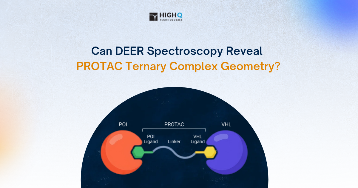
A PROTAC ternary complex is an assembly of three components: a target protein, a PROTAC molecule and an E3 ubiquitin ligase. A PROTAC is a bifunctional molecule consisting of two binding groups connected by a chemical linker:
- Target-binding group (warhead): Binds the protein selected for degradation.
- E3 ligase-binding group: Recruits an E3 ubiquitin ligase, which helps transfer ubiquitin onto the target protein.
- Chemical linker: Connects the two binding groups and influences how the target and E3 ligase are positioned relative to one another.
The PROTAC binds to both the target protein and the E3 ligase, bringing them together to form a ternary complex. The E3 ligase facilitates the attachment of ubiquitin molecules to the target protein. Once sufficiently ubiquitinated, the target is recognized and degraded by the cell's proteasome. Following degradation, the PROTAC can be released and potentially participate in another degradation cycle, allowing it to act catalytically.
How Does Ternary-Complex Geometry Affect Targeted Protein Degradation?
The formation of a stable ternary complex is an important step in PROTAC-mediated degradation. Nevertheless, complex stability alone does not determine degradation efficiency. The interactions between the target protein and E3 ligase also contribute to ternary-complex formation and can influence how effectively the complex supports ubiquitination.
These interactions are described by cooperativity. Positive cooperativity occurs when binding of the PROTAC to one protein enhances its interaction with the second, often through favourable protein–protein interactions (PPIs) at the interface. Negative cooperativity occurs when these interactions make ternary-complex formation less favourable. Noteworthy, the relative positioning of the target protein and E3 ligase can influence whether the complex supports productive ubiquitination. The E3 ligase must be positioned so that ubiquitin can be transferred to accessible lysine residues on the target. Consequently, two PROTACs can form ternary complexes with comparable binding properties but differ in their ability to promote target degradation.
This relationship was investigated by Wurz and colleagues at Amgen in a 2023 study published in Nature Communications. Using VHL-recruiting PROTACs targeting SMARCA2 and BRD4, the researchers found that ternary-complex binding affinity and cooperativity correlated with degradation potency and initial degradation rates. Their findings also showed that changes to PROTAC linker structure and ligand orientation influenced ternary-complex formation and degradation activity.
Another important consideration is conformational heterogeneity. PROTAC-induced ternary complexes do not necessarily adopt one fixed structure. The flexibility of the linker and the interactions between the target and E3 ligase can give rise to multiple possible arrangements, some of which may be more favorable for ubiquitination than others. Bai and colleagues explored this challenge in a 2021 study published in the Journal of Chemical Information and Modeling. Using computational modelling, they demonstrated that PROTAC activity is better understood by considering an ensemble of possible ternary-complex structures rather than a single static conformation. They also showed that the same PROTAC could recruit structurally related proteins through different binding modes.
These findings highlight why understanding ternary-complex geometry and conformational behavior matters in PROTAC design. Measuring how the target and E3 ligase are positioned relative to one another, and how their arrangement varies, could provide additional insight into why some degraders are more effective than others.
Techniques Used to Characterize PROTAC Ternary Complexes
Several biophysical and structural techniques are used to investigate PROTAC-induced ternary complexes. Each method provides different information about binding, complex formation and structural organization.
Surface Plasmon Resonance (SPR) and Biolayer Interferometry (BLI)
SPR and BLI measure molecular interactions in real time, providing information about binding affinity and association and dissociation kinetics. In PROTAC research, they can be used to characterize binary interactions and ternary-complex formation, including the effects of linker design and cooperativity.
However, these measurements do not directly provide distance distributions between selected regions of the interacting proteins.
Isothermal Titration Calorimetry (ITC)
ITC measures the heat released or absorbed during molecular binding. It provides information about binding affinity, stoichiometry, and thermodynamic parameters, including enthalpy and entropy. It can also be used to investigate ternary-complex formation and cooperativity without requiring fluorescent labels or surface immobilization.
However, ITC does not directly reveal the relative positioning or conformational heterogeneity of the proteins.
Fluorescence Resonance Energy Transfer (FRET)
FRET-based assays investigate molecular proximity through energy transfer between fluorescent donor and acceptor labels. Approaches such as TR-FRET and NanoBRET are used to monitor PROTAC-induced interactions, with NanoBRET enabling measurements in living cells.
FRET can provide distance-sensitive information, but its interpretation depends on label placement, orientation and the properties of the fluorophores. A single FRET measurement does not establish the full structural arrangement of a ternary complex.
X-ray Crystallography (XRC) and Cryo-Electron Microscopy (Cryo-EM)
X-ray crystallography can provide atomic-level structural information about PROTAC-induced ternary complexes, including protein–protein interfaces and ligand-binding interactions. However, obtaining suitable crystals can be challenging, and the resulting structure may not capture the full range of conformations present in solution.
Cryo-EM can provide structural information about larger protein complexes and, through classification, help identify different structural states. However, the size, flexibility and conformational heterogeneity of some PROTAC-induced complexes can make structural determination challenging.
What Could Distance Distributions from Pulsed Dipolar EPR Spectroscopy Add?
While existing techniques provide information about binding, interaction kinetics, thermodynamics and structural architecture, they do not always show how specific regions of the target protein and E3 ligase are positioned relative to one another within a ternary complex.
Pulsed dipolar electron paramagnetic resonance (EPR) spectroscopy, particularly double electron–electron resonance (DEER), could provide a complementary measurement. DEER, also known as PELDOR, measures the magnetic dipolar interaction between two unpaired electron spins. In protein research, these spins are commonly introduced through site-directed spin labelling (SDSL), in which spin labels are attached at selected positions, often using engineered cysteine residues.
DEER measurements produce interspin distance distributions that provide information about protein conformations and structural heterogeneity. When labels are introduced on different protein components, the technique can also measure distances between them, providing experimental constraints on their relative positioning. Typical distance ranges for nitroxide-labelled proteins are approximately 1.5–8 nm, with longer distances accessible under suitable experimental conditions.
How Could DEER Be Applied to PROTAC Ternary Complexes?
One possible approach would be to introduce spin labels at selected sites on the target protein and the E3 ligase complex. For example, in a PROTAC system recruiting a target protein to a VHL- or CRBN-containing E3 ligase complex, labels could be placed at defined positions on the target and the corresponding E3 ligase substrate receptor. DEER measurements could then provide distance distributions between these sites in the assembled ternary complex. Researchers could investigate the effects of different experimental conditions, including:
- Formation of binary versus ternary complexes.
- Different PROTAC linker designs.
- Different E3 ligase-recruiting elements.
- Alternative target–E3 ligase combinations.
Different PROTACs recruiting the same target and E3 ligase could produce distinct distance distributions, reflecting differences in the relative positioning and conformational heterogeneity of the complex. These measurements could provide structural information to interpret alongside ternary-complex binding and degradation data. Careful selection of spin-labelling sites would be essential to avoid disrupting protein structure, binding interfaces or ternary-complex formation.
Accessing DEER Measurements for PROTAC Research
DEER measurements can be performed using an in-house pulsed EPR spectrometer or through an external measurement service. However, sample volume, measurement time, and the need for an EPR specialist have historically kept the technique out of drug discovery groups. High Q Technologies' FATHOM platform addresses these barriers through an automated, quantum-enabled pulsed EPR workflow. Its superconducting quantum-sensing resonator enables sensitive distance-distribution measurements using small sample volumes. FATHOM® supports automated sample loading, calibration, measurement and distance analysis, handling up to five samples per cartridge, with sample volumes as low as 3.5 µL per sample. The system also operates without the need for liquid cryogens.
For researchers who do not have access to an in-house EPR instrument, High Q's Sample Measurement Service provides customized measurements using FATHOM®, together with detailed reports and measurement data.
If you are considering DEER spectroscopy for a PROTAC ternary-complex study, speak with an Applications Scientist to discuss your target proteins, spin-labelling strategystrategy, and experimental requirements.
Key takeaways
- Ternary-complex formation does not necessarily lead to efficient protein degradation. The relative positioning and conformation of the target and E3 ligase also influence ubiquitination.
- SPR, BLI, ITC, FRET, X-ray crystallography and cryo-EM provide complementary information on binding and complex structure.
- DEER spectroscopy measures distance distributions between selected spin-labelled sites, providing insight into protein positioning and conformational heterogeneity.
- Distance measurements alongside structural and functional data could help explain differences in PROTAC degradation activity.
- High Q Technologies' FATHOM® platform and Sample Measurement Service provide access to pulsed EPR measurements for protein research.
Frequently asked questions
Does ternary complex formation always lead to degradation?
No. A degrader can assemble a stable, measurable ternary complex and still degrade nothing, because ubiquitin transfer also requires a surface lysine on the target to sit within reach of the ligase active site. This is why potency often fails to track binding affinity across a linker series, and why teams measure ubiquitination and cellular degradation separately. Structural information about the complex helps explain which complexes are productive.
What is cooperativity in a PROTAC ternary complex?
Cooperativity describes whether the presence of one protein makes the degrader bind the other more tightly or less tightly, expressed as the ratio of binary to ternary dissociation constants. Positive cooperativity indicates favorable contacts between the target and the E3 ligase themselves. Wurz and colleagues reported in Nature Communications in 2023 that cooperativity, together with ternary binding affinity, correlated with degradation potency across VHL-recruiting degraders of SMARCA2 and BRD4.
Can you measure PROTAC ternary complexes in live cells?
Partly. Cellular proximity assays such as NanoBRET report whether the target and the ligase come together inside cells, and degradation assays report the outcome. What these do not give is the arrangement of the complex. DEER is a frozen-sample method run on purified, spin-labelled components, so it addresses geometry in a reconstituted system rather than in cells.
How is a molecular glue different from a PROTAC?
A molecular glue is a single small molecule that stabilizes a direct interface between a target protein and an E3 ligase, while a PROTAC is a bifunctional molecule with two binding ends joined by a linker. Glues are smaller and more similar to drugs, and their activity depends almost entirely on the protein-protein interface, since there is no linker to tune. Both work through the same ubiquitin-proteasome pathway.
Do you need to mutate cysteines to spin label a protein?
Usually yes. The standard nitroxide label reacts with a cysteine thiol, so a cysteine is introduced by site-directed mutagenesis at the position you want to investigate, and native surface cysteines are mutated away first so the label specifically attaches at the artificially introduced cysteine. A ternary complex therefore needs two separate single-cysteine constructs, one for the target and one for the ligase. Alternative labeling chemistries exist where cysteine engineering can be avoided.
References
- Tsai, J. M. et al. Targeted protein degradation: from mechanisms to clinic. Nature Reviews Molecular Cell Biology. 2024;25:740–757. https://doi.org/10.1038/s41580-024-00729-9
- Casement, R., Bond, A., Craigon, C. and Ciulli, A. Mechanistic and Structural Features of PROTAC Ternary Complexes. Methods in Molecular Biology. 2021;2365:79–113. https://doi.org/10.1007/978-1-0716-1665-9_5
- Affinity and cooperativity modulate ternary complex formation to drive targeted protein degradation. Nature Communications. 2023. https://doi.org/10.1038/s41467-023-39904-5
- Ponzo, I. et al. Proximity Binding Assay for PROTAC Ternary Complex Analysis. ACS Sensors. 2026;11(5):3876–3888. https://doi.org/10.1021/acssensors.5c04944
- A real-time FRET ubiquitin transfer assay for quantitative characterization of ternary complexes in targeted protein degradation. Cell Reports Methods. 2026. https://doi.org/10.1016/j.crmeth.2026.101505
- Tessmer, M. H. and Stoll, S. Protein Modeling with DEER Spectroscopy. Annual Review of Biophysics. 2025;54:35–57. https://doi.org/10.1146/annurev-biophys-030524-013431
- Benchmark Test and Guidelines for DEER/PELDOR Experiments on Nitroxide-Labeled Biomolecules. Journal of the American Chemical Society. 2021. https://doi.org/10.1021/jacs.1c07371
- Cross-validation of distance measurements in proteins by PELDOR/DEER and single-molecule FRET. Nature Communications. 2022. https://doi.org/10.1038/s41467-022-31945-6
- Nan Bai, Sven A. Miller, Grigorii V. Andrianov, Max Yates, Palani Kirubakaran, John Karanicolas; Rationalizing PROTAC-Mediated Ternary Complex Formation Using Rosetta. J. Chem. Inf. Model. 22 March 2021; 61 (3): 1368–1382. https://doi.org/10.1021/acs.jcim.0c01451
- Bai N, Miller SA, Andrianov GV, Yates M, Kirubakaran P, Karanicolas J. Rationalizing PROTAC-mediated ternary complex formation using Rosetta. J Chem Inf Model. 2021 Mar 22;61(3):1368-1382. doi: 10.1021/acs.jcim.0c01451. PMID: 33625214; PMCID: PMC8866032.