Protein dimerization occurs when two protein molecules associate to form a dimer. A crystal structure can reveal how two protein molecules fit together, identifying an interface that may influence protein function or drug discovery. However, a dimer observed in a crystal does not necessarily represent the dominant assembly in solution. Crystal packing can stabilize contacts that are weak, transient, or absent under physiologically relevant conditions. For pharmaceutical researchers, this distinction matters because a proposed dimer interface may form a binding site, regulate protein activity, or provide a target for small molecules. Before using the structure to guide drug design, researchers need to establish whether the protein associates in solution and whether the arrangement of its subunits is consistent with the proposed model.
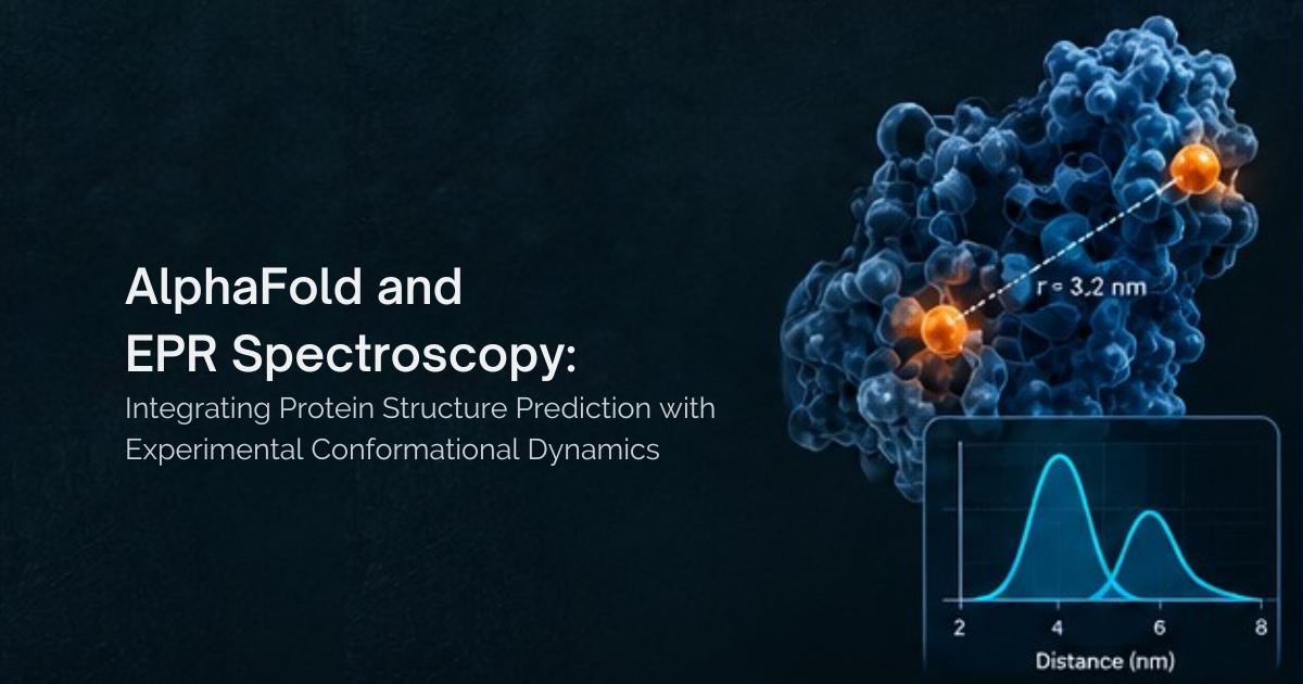
Electron paramagnetic resonance (EPR) spectroscopy provides a way to investigate protein association and structure through measurements of spin-label interactions and distances between selected sites. In particular, site-directed spin labeling combined with pulsed dipolar EPR, commonly called double electron–electron resonance (DEER) or PELDOR, can test whether measured distances support a proposed interface. When combined with complementary solution methods, these measurements provide experimental constraints for evaluating protein assemblies.
Why a Crystal Structure May Not Reflect Protein Dimerization in Solution
Proteins can form dimers through specific biological interactions, but they can also make contacts favoured by the high local concentrations and packing constraints of crystallization. A crystal structure alone cannot always distinguish between these possibilities. The solution state may be more complex than a single structure suggests. Some proteins exist in a concentration-dependent equilibrium between monomers and dimers, while others populate multiple oligomeric states or undergo transient association. Protein concentration, buffer composition, pH, temperature, and ligand binding can all influence the observed behaviour. Researchers therefore need to establish whether a dimer forms under relevant solution conditions, whether its subunit arrangement agrees with the proposed structure, and whether the assembly has a biological or functional role. These are related but distinct questions that may require complementary experiments.
How Researchers Test Protein Dimerization in Solution
Size-exclusion chromatography coupled to multi-angle light scattering (SEC-MALS) estimates the molar mass of species separated by chromatography, helping distinguish monomers from dimers when the species are sufficiently resolved and stable during measurement. Analytical ultracentrifugation provides complementary information about protein association in solution and, with appropriate experimental design and analysis, can help characterize concentration-dependent equilibria. Native mass spectrometry may also provide evidence of oligomeric stoichiometry when suitable for the protein complex and experimental conditions. Each method has limitations. For weak or rapidly exchanging complexes, chromatography can alter the apparent distribution of species through dilution or dissociation. Concentration-series experiments can help reveal reversible association, but concentration dependence alone does not establish that a specific interface is responsible. Nonspecific interactions, aggregation, and method-specific effects must also be considered. These measurements help characterize which oligomeric species are present. EPR can add a different type of evidence by testing whether distances between selected sites are consistent with a proposed subunit arrangement.
How Electron Paramagnetic Resonance Investigates Protein Dimerization
The information obtained from EPR depends on the method, labeling strategy, and question being investigated. Continuous-wave EPR can provide information about spin-label mobility and interactions, while DEER measures distances and distance distributions between spin labels.
Investigating Association with Continuous-Wave EPR
Continuous-wave EPR (CW-EPR) can report local spin-label mobility and spin–spin interactions. When labeled protein molecules associate, interactions between their spin labels may alter the observed spectral line shape. Such changes can provide evidence of association when the labeling design and experimental conditions make the effects interpretable. Under suitable conditions, an appropriate labeling strategy and validated spectral analysis may also help estimate the relative populations of associated and unassociated species. However, CW-EPR does not automatically provide quantitative monomer and dimer fractions. Spectral changes can also arise from differences in local mobility or environment, so appropriate controls are essential.
Testing a Symmetric Homodimer Interface with DEER
For a symmetric homodimer, a common starting strategy is to introduce a spin label at the same selected residue in each subunit. The resulting intersubunit distance can be compared with the distance predicted by the crystal-derived model. Label positions should be selected to distinguish between the proposed interface and plausible alternative arrangements. The chosen residues should also permit effective labeling without substantially disrupting the interaction. Label mobility, labeling efficiency, and protein flexibility must be considered when interpreting the measurements. One carefully selected labeling position can provide an initial test of a predicted distance, but agreement with a single distance does not uniquely establish interface geometry. Multiple independent labeling positions generally provide stronger structural constraints and help distinguish competing models. The number needed depends on the complexity of the assembly, the differences between candidate structures, and the quality of the data.
Symmetric oligomers also require care during DEER analysis because multiple spin pairs may contribute to the measured signal, depending on the labeling pattern and oligomeric arrangement. Appropriate sample characterization and analysis of potential multi-spin effects are therefore important. The DEER/PELDOR experimental guidelines discuss key considerations for labeling, sample preparation, measurement quality, and data interpretation. Importantly, DEER measures distances and distance distributions between spin labels; it does not independently establish what fraction of the protein exists as a dimer. Combining distance measurements with independent oligomeric-state characterization provides a more complete assessment.
Distinguishing a Crystal-Packing Contact from a Weak Dimer
Researchers can investigate a proposed interface by examining the protein over a range of concentrations while keeping other experimental conditions as consistent as possible. If a reversible monomer–dimer equilibrium is present, the relative populations may shift with concentration. These observations can be compared with EPR results and measurements from an independent solution method. A concentration-dependent change in an EPR spectrum or distance signal may support an association model, but it is not definitive on its own. Concentration can also influence nonspecific interactions, aggregation, and the intermolecular background in DEER measurements.
Sample preparation is another consideration because DEER measurements are commonly performed on frozen samples. Freezing and cryoprotectant conditions can influence weak protein–protein interactions, so the observed distribution should not automatically be assumed to represent the equilibrium present in liquid solution. Freezing does not necessarily invalidate a measurement; potential effects need to be evaluated for the particular protein and interaction. Appropriate controls and independent evidence of oligomeric state strengthen the interpretation. See Schmidt et al., Submillisecond Freezing Permits Cryoprotectant-Free EPR Double Electron–Electron Resonance Spectroscopy, for research on sample preparation for DEER spectroscopy.
Testing Interface Geometry and Structural Heterogeneity
Once there is evidence that the protein associates in solution, DEER spectroscopy can help test whether the relative arrangement of the subunits is consistent with the crystal-derived model. Researchers compare measured distance distributions with distances predicted from the structure, accounting for the accessible conformations of the spin labels. Agreement across multiple independent distances supports the compatibility of the model with the measurements. Disagreement may indicate that the crystal structure does not capture the dominant solution arrangement, that the protein samples several conformations, or that experimental assumptions need to be revisited. A discrepancy does not automatically invalidate the crystal structure; it may reveal that the structure represents one state within a broader ensemble. DEER spectroscopy can also reveal structural heterogeneity when distinct conformations are resolved by the experiment and supported by reliable analysis. Comparing measurements in the presence and absence of a ligand can further test whether binding changes the distances or conformational distributions associated with the dimer. Functional assays are still needed to establish the biological consequences of such changes.
Choosing Complementary Methods
The most useful combination of techniques depends on the uncertainty researchers need to resolve. SEC-MALS, analytical ultracentrifugation, and, where suitable, native mass spectrometry can help characterize oligomeric state. CW-EPR can provide information about spin-label mobility and interactions, while DEER adds distance restraints for testing a proposed subunit arrangement. For a weak or reversible dimer, concentration-dependent measurements are particularly valuable. For a structure intended to guide drug design, multiple independent distances can help distinguish the proposed interface from alternative arrangements. Across these experiments, sample quality, labeling efficiency, removal of free spin label where appropriate, suitable data analysis, and reproducibility remain important. These considerations are addressed in the DEER/PELDOR benchmark and guidelines paper.
Why Protein Dimerization Matters for Drug Discovery
An unvalidated protein interface can introduce uncertainty into decisions about binding sites, allosteric regulation, protein–protein interactions, and compound design. The implications depend on the role of the assembly in the target's biology. For example, a small molecule may be designed to disrupt a proposed protein–protein interface. If the dimer is not appreciably populated under the relevant solution conditions, researchers may need to reconsider the proposed binding mechanism or determine whether the interaction occurs only under specific conditions. Oligomeric-state measurements can help establish which assemblies are present, while DEER distance restraints can test whether the proposed interface geometry is consistent with experimental observations. Neither measurement alone establishes that a compound will be effective, but together they can help assess the structural assumptions underlying a drug-discovery strategy. The practical value is greater confidence in deciding which structural models warrant further investigation, which experiments are needed to resolve uncertainty, and whether a proposed mechanism remains consistent with the available evidence.
Testing Protein Models with High Q Technologies
High Q Technologies develops EPR spectroscopy systems for investigating protein structure and dynamics. For researchers studying protein dimerization, EPR measurements can complement established structural and biophysical methods by providing information about spin-label interactions and distances between selected sites.
The goal is to connect proposed protein interfaces with experimental evidence under defined conditions. To explore how EPR spectroscopy can support protein characterization and structural studies, visit High Q Technologies.
References
- Marciano, S. et al. (2022). “Protein quaternary structures in solution are a mixture of multiple forms.” Chemical Science. https://doi.org/10.1039/D2SC02794A
- Schiemann, O. et al. (2021). “Benchmark Test and Guidelines for DEER/PELDOR Experiments on Nitroxide-Labeled Biomolecules.” Journal of the American Chemical Society, 143, 17875–17890. https://doi.org/10.1021/jacs.1c07371
- Schmidt, M. J. et al. (2020). “Submillisecond Freezing Permits Cryoprotectant-Free EPR Double Electron–Electron Resonance Spectroscopy.” ChemPhysChem. https://doi.org/10.1002/cphc.202000312