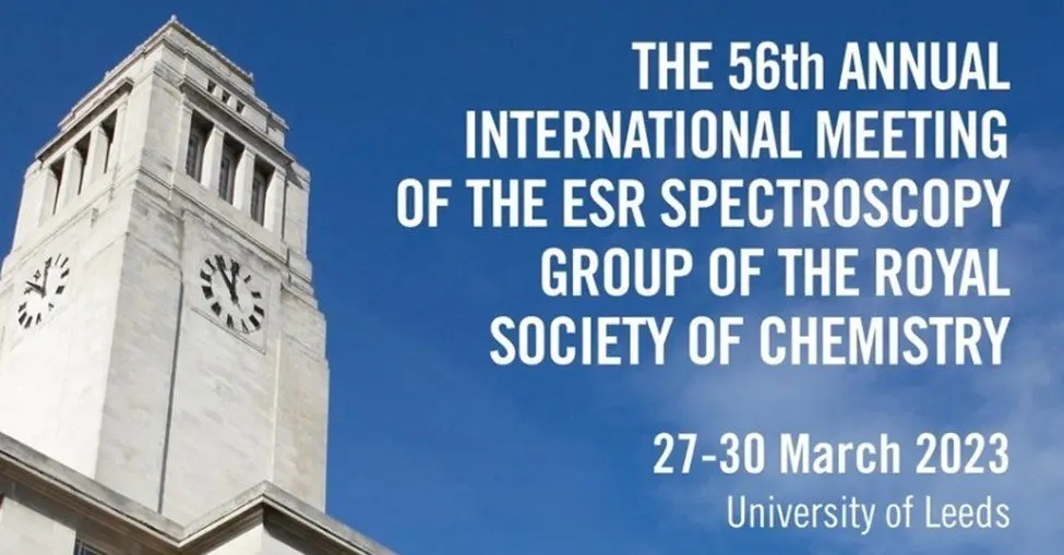
In my inaugural post of Insights, I took a bird’s-eye view of how EPR can fit into the broad and diverse landscape of biophysics – an apt reflection after a meeting of the Biophysical Society. This past week I ventured to Leeds to join the Royal Society of Chemistry’s ESR meeting (EPR and ESR are interchangeable) and for a meeting of such focused subject matter, this reflection will appropriately take a more focused approach.
However, even in such an EPR-centric meeting, the presentations and subject matter were incredibly varied. I witnessed work that ranged from materials science, to software, hardware, and method development, to quantum computing. While the scientific brilliance was clear and evident across the many excellent talks, given that I have “EPR for Protein Dynamics” printed on my business cards, I was particularly impressed with the structural biology that was presented, and am only further convinced that EPR has a bigger part to play within the scientific community.
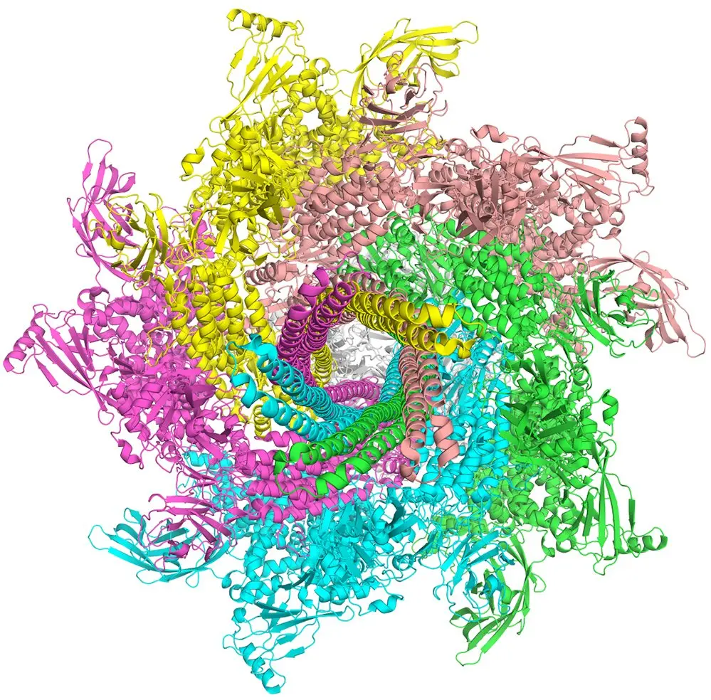
Prof. Enrica Bordignon (Université de Genève) exemplified this notion early on in the conference. Her presentation covered many of the virtues of EPR – beginning with the subject of her investigations, Tc toxin, a class of large protein complexes weighing in around 1.7 MDa. Such a massive unit is quite inaccessible via NMR, and crystallization would be complicated, to say the least. Bordignon’s EPR work – in which EPR-based distance and dynamics measurements were used to elucidate the kinetics of the toxin’s mechanics – was done in complement to Cryo-EM and smFRET to identify discrete intermediate conformations of the protein assembly. Her presented body of work is an excellent example of what I envision for EPR – a powerful resolver of protein conformations, dynamics, and even kinetics, working in tandem with other techniques to gain a complete picture of vital biological and physiological systems.
In a similar vein, Dr. Thomas Schmidt’s (NIH, NIDDK) projects involved GPCRs (G-protein coupled receptors), an important class of proteins that mediate cellular responses to stimuli and present an attractive target for drug development. Specifically, Schmidt used a global analysis method to elucidate the equilibrium of a monomer/dimer GPCR system governed by ligand interactions. His work reiterated the ability of EPR to gather deep insights into the whole of a biological process – investigating structure, dynamics, conformations, and kinetics all at once. On a technical level, the experimentalist in me appreciated a clever scheme in which he filtered out contributions from the monomers or dimers in his distance measurements by manipulating the differences in relaxation rates of the two species – an idea that may prove extremely valuable in efforts to deconvolute complicated multimodal distance distributions.
Finally, the work of Prof. Bela Bode (University of St. Andrews) pushed the boundaries of sensitivity in EPR measurements. In short, he showed that such measurements could be achieved on protein systems with concentrations as low as 50 nM. Typical EPR spectroscopy uses protein concentrations in the 100 μM range – 3-4 orders of magnitude higher. Lowering that limit is hugely important when transitioning from establishing methodologies on model proteins to collecting data on real, biologically relevant systems. Model systems are often chosen for their good behavior and ease of manipulation, such that generating 100 μM of such a protein can be a routine task. Not all – few, in fact – proteins are so polite, and lower the threshold of protein necessary will make previously prohibitively tricky macromolecules accessible by EPR.

While I only have the wordcount to discuss a few cases here, the sum of work presented at RSC paints a bright picture for the field. The work can be hard, frustrating, and oftentimes slower than we’d like, but through the diligence of talented and determined scientists, EPR as a technique has seen monumental growth since its inception. It’s a testament to the community to see the work presented at conferences like RSC that, year after year, constantly push the state of the art forward to places previously unforeseen. I’m excited to see the role that I, and especially High Q, have to play in the coming years. And even more than that – I’m excited to share it with the rest of you. In the meantime, there’s no rest for the weary – the High Q Crew is headed back to San Diego next week to look into the intersection of EPR and drug discovery at Drug Discovery Chemistry 2023. See you in California!