
Join us in Snowbird, Utah, from August 02–06, 2026 for the 65th Annual Rocky Mountain Conference on Magnetic Resonance (RMCMR)
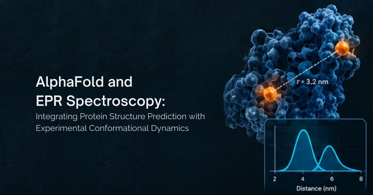
Few tools have changed structural biology as quickly as AlphaFold2. Within a few years of its release, AlphaFold2 had become an important starting point for structural studies of many proteins. However, transporters, receptors, channels and many enzymes function through conformational changes, and ligand binding can shift proteins between different structural states. A single predicted model, however confident, represents one structural state rather than the full range of conformations a protein may populate. This is where Electron Paramagnetic Resonance (EPR) spectroscopy comes in.
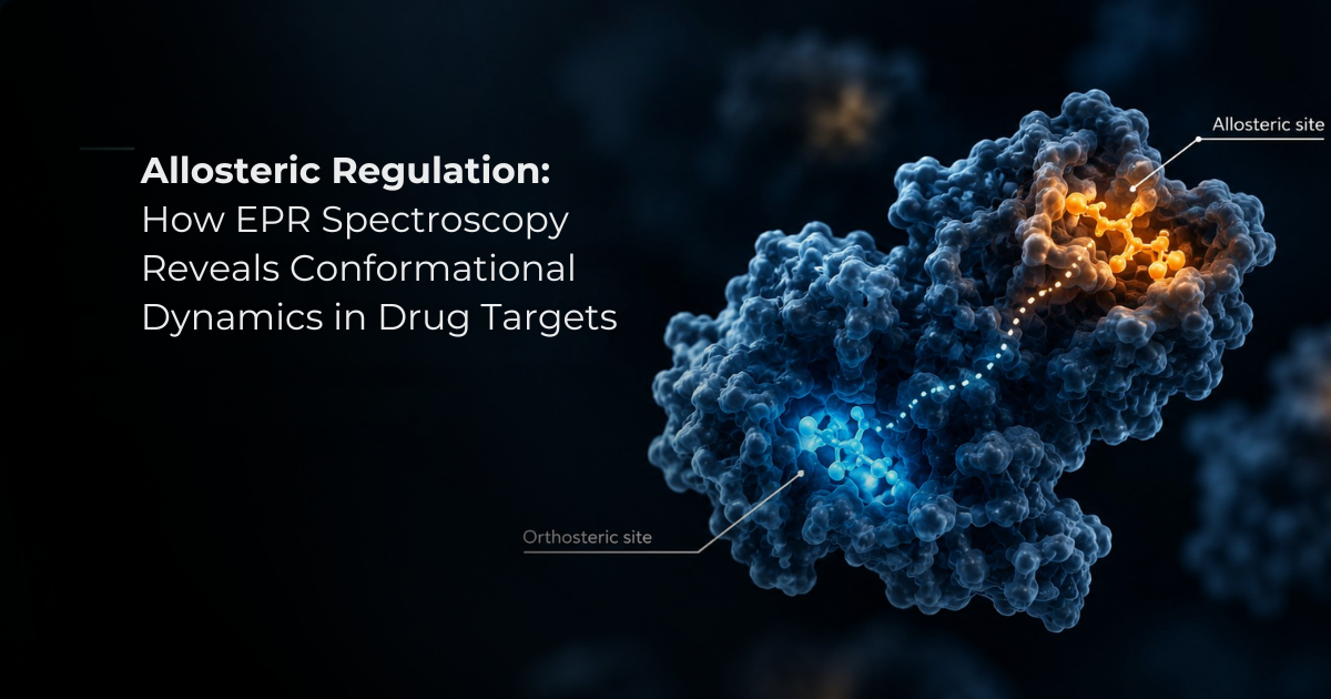
Allosteric regulation is a fundamental mechanism of cellular control, enabling proteins to respond to signals at one site and translate them into changes in activity at another, often distant, site. Proteins regulated in this way, known as allosteric proteins, are involved in a wide range of biological processes, including enzyme regulation, signal transduction, cell adhesion and transcription.
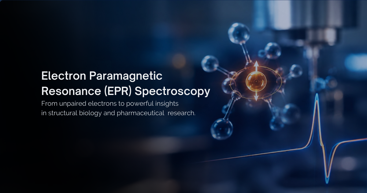
Electron paramagnetic resonance spectroscopy is a powerful way to study materials, molecules, and systems with unpaired electrons. It is often called EPR, ESR, electron spin resonance spectroscopy, or spin resonance spectroscopy.
Ready to see FATHOM in action?
Our applications specialists will walk you through a demo, answer technical questions, and help you assess fit for your specific research program.